Phyre Home Retrieve Phyre Job Id
Emaill.a.kelley@imperial.ac.uk
DescriptionP0AEL0
DateThu Jan 5 11:23:36 GMT 2012
Unique Job ID424399b82e571ae4

Summary 

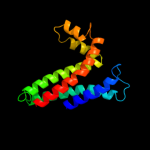
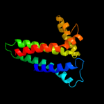
Top model
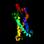
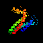
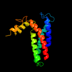
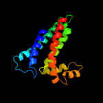
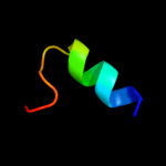
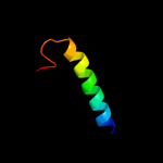
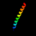
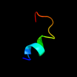
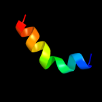
Image coloured by rainbow N → C terminus
Model (left) based on template d1kqfc_
Top template information
Fold:Heme-binding four-helical bundle
Superfamily:Transmembrane di-heme cytochromes
Family:Formate dehydrogenase N, cytochrome (gamma) subunit
Confidence and coverage
Confidence:100.0% Coverage: 99%
209 residues ( 99% of your sequence) have been modelled with 100.0% confidence by the single highest scoring template.
3D viewing
Interactive 3D view in Jmol

Sequence analysis 

Secondary structure and disorder prediction 

   1........10.........20.........30.........40.........50.........60
Sequence  MKRRDTIVRYTAPERINHWITAFCFILAAVSGLGFLFPSFNWLMQIMGTPQLARILHPFV
Secondary structure 


SS confidence 


Disorder  ?????


Disorder confidence 


   .........70.........80.........90.........100.........110.........120
Sequence  GVVMFASFIIMFFRYWHHNLINRDDIFWAKNIRKIVVNEEVGDTGRYNFGQKCVFWAAII
Secondary structure 


SS confidence 


Disorder 


?


??


Disorder confidence 


   .........130.........140.........150.........160.........170.........180
Sequence  FLVLLLVSGVIIWRPYFAPAFSIPVIRFALMLHSFAAVALIVVIMVHIYAALWVKGTITA
Secondary structure 


SS confidence 


Disorder 


????


Disorder confidence 


   .........190.........200.........210.
Sequence  MVEGWVTSAWAKKHHPRWYREVRKTTEKKAE
Secondary structure 


SS confidence 


Disorder 


????????
Disorder confidence 


Confidence Key
High(9)                    Low (0)
?Disordered
Alpha helix
Beta strand

Domain analysis 

Hover over an aligned region to see model and summary info

Please note, only up to the top 20 hits are modelled to reduce computer load

RankAligned region

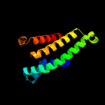
PDB 1kqf chain C

3D model

Region: 2 - 210
Aligned: 209
Modelled: 209
Confidence: 100.0%
Identity: 48%
Fold: Heme-binding four-helical bundle
Superfamily: Transmembrane di-heme cytochromes
Family: Formate dehydrogenase N, cytochrome (gamma) subunit

Phyre2

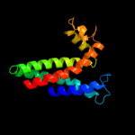
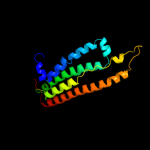
PDB 2qjk chain M

3D model

Region: 20 - 171
Aligned: 152
Modelled: 152
Confidence: 98.2%
Identity: 17%
PDB header:electron transport
Chain: M: PDB Molecule:cytochrome b;
PDBTitle: crystal structure analysis of mutant rhodobacter2 sphaeroides bc1 with stigmatellin and antimycin

Phyre2

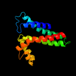
PDB 1q90 chain B

3D model

Region: 20 - 171
Aligned: 152
Modelled: 152
Confidence: 98.1%
Identity: 19%
Fold: Heme-binding four-helical bundle
Superfamily: Transmembrane di-heme cytochromes
Family: Cytochrome b of cytochrome bc1 complex (Ubiquinol-cytochrome c reductase)

Phyre2

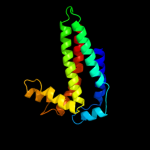
PDB 3cx5 chain C domain 2

3D model

Region: 20 - 171
Aligned: 152
Modelled: 152
Confidence: 98.1%
Identity: 14%
Fold: Heme-binding four-helical bundle
Superfamily: Transmembrane di-heme cytochromes
Family: Cytochrome b of cytochrome bc1 complex (Ubiquinol-cytochrome c reductase)

Phyre2

PDB 3cx5 chain N

3D model

Region: 20 - 171
Aligned: 152
Modelled: 152
Confidence: 98.1%
Identity: 14%
PDB header:oxidoreductase
Chain: N: PDB Molecule:cytochrome b;
PDBTitle: structure of complex iii with bound cytochrome c in reduced2 state and definition of a minimal core interface for3 electron transfer.

Phyre2

PDB 3cwb chain C

3D model

Region: 20 - 171
Aligned: 151
Modelled: 152
Confidence: 98.1%
Identity: 14%
PDB header:oxidoreductase
Chain: C: PDB Molecule:cytochrome b;
PDBTitle: chicken cytochrome bc1 complex inhibited by an iodinated analogue of2 the polyketide crocacin-d

Phyre2

PDB 1bcc chain C domain 3

3D model

Region: 20 - 171
Aligned: 151
Modelled: 152
Confidence: 98.0%
Identity: 14%
Fold: Heme-binding four-helical bundle
Superfamily: Transmembrane di-heme cytochromes
Family: Cytochrome b of cytochrome bc1 complex (Ubiquinol-cytochrome c reductase)

Phyre2

PDB 1ppj chain C domain 2

3D model

Region: 20 - 171
Aligned: 151
Modelled: 152
Confidence: 98.0%
Identity: 13%
Fold: Heme-binding four-helical bundle
Superfamily: Transmembrane di-heme cytochromes
Family: Cytochrome b of cytochrome bc1 complex (Ubiquinol-cytochrome c reductase)

Phyre2

PDB 2e74 chain A domain 1

3D model

Region: 20 - 171
Aligned: 152
Modelled: 152
Confidence: 98.0%
Identity: 18%
Fold: Heme-binding four-helical bundle
Superfamily: Transmembrane di-heme cytochromes
Family: Cytochrome b of cytochrome bc1 complex (Ubiquinol-cytochrome c reductase)

Phyre2

PDB 1y5i chain C domain 1

3D model

Region: 16 - 165
Aligned: 115
Modelled: 124
Confidence: 42.5%
Identity: 17%
Fold: Heme-binding four-helical bundle
Superfamily: Respiratory nitrate reductase 1 gamma chain
Family: Respiratory nitrate reductase 1 gamma chain

Phyre2

PDB 3mk7 chain K

3D model

Region: 1 - 191
Aligned: 168
Modelled: 169
Confidence: 33.4%
Identity: 14%
PDB header:oxidoreductase
Chain: K: PDB Molecule:cytochrome c oxidase, cbb3-type, subunit n;
PDBTitle: the structure of cbb3 cytochrome oxidase

Phyre2

PDB 2kyg chain C

3D model

Region: 183 - 205
Aligned: 22
Modelled: 23
Confidence: 33.3%
Identity: 27%
PDB header:protein binding
Chain: C: PDB Molecule:protein cbfa2t1;
PDBTitle: structure of the aml1-eto nervy domain - pka(riia) complex and its2 contribution to aml1-eto activity

Phyre2

PDB 2lev chain A

3D model

Region: 196 - 211
Aligned: 16
Modelled: 16
Confidence: 31.3%
Identity: 38%
PDB header:transcription regulator/dna
Chain: A: PDB Molecule:ler;
PDBTitle: structure of the dna complex of the c-terminal domain of ler

Phyre2

PDB 3kmp chain A

3D model

Region: 184 - 211
Aligned: 28
Modelled: 28
Confidence: 14.0%
Identity: 21%
PDB header:transcription regulator/dna
Chain: A: PDB Molecule:smad1-mh1;
PDBTitle: crystal structure of smad1-mh1/dna complex

Phyre2

PDB 1m56 chain D

3D model

Region: 1 - 34
Aligned: 34
Modelled: 34
Confidence: 13.4%
Identity: 12%
Fold: Single transmembrane helix
Superfamily: Bacterial aa3 type cytochrome c oxidase subunit IV
Family: Bacterial aa3 type cytochrome c oxidase subunit IV

Phyre2

PDB 1hnr chain A

3D model

Region: 196 - 211
Aligned: 16
Modelled: 16
Confidence: 9.7%
Identity: 6%
Fold: H-NS histone-like proteins
Superfamily: H-NS histone-like proteins
Family: H-NS histone-like proteins

Phyre2

PDB 2od6 chain A domain 1

3D model

Region: 188 - 206
Aligned: 19
Modelled: 19
Confidence: 7.0%
Identity: 21%
Fold: Ferredoxin-like
Superfamily: Dimeric alpha+beta barrel
Family: Marine metagenome family DABB1

Phyre2

PDB 2l4g chain A

3D model

Region: 168 - 188
Aligned: 21
Modelled: 17
Confidence: 6.0%
Identity: 14%
PDB header:viral protein
Chain: A: PDB Molecule:haemagglutinin;
PDBTitle: influenza haemagglutinin fusion peptide mutant g13a

Phyre2

PDB 1fcu chain A

3D model

Region: 187 - 210
Aligned: 24
Modelled: 24
Confidence: 6.0%
Identity: 17%
PDB header:hydrolase
Chain: A: PDB Molecule:hyaluronoglucosaminidase;
PDBTitle: crystal structure (trigonal) of bee venom hyaluronidase

Phyre2

PDB 1rrz chain A

3D model

Region: 182 - 201
Aligned: 15
Modelled: 20
Confidence: 5.9%
Identity: 27%
Fold: Spectrin repeat-like
Superfamily: Glycogen synthesis protein GlgS
Family: Glycogen synthesis protein GlgS

Phyre2
1

d1kqfc_
2

c2qjkM_
3

d1q90b_
4

d3cx5c2
5

c3cx5N_
6

c3cwbC_
7

d1bccc3
8

d1ppjc2
9

d2e74a1
10

d1y5ic1
11

c3mk7K_
12

c2kygC_
13

c2levA_
14

c3kmpA_
15

d1m56d_
16

d1hnra_
17

d2od6a1
18

c2l4gA_
19

c1fcuA_
20

d1rrza_
21


22


23


24


25


26


27


28


Detailed template information 

#
Template Alignment Coverage3D Model Confidence
% i.d. Template Information
1d1kqfc_


100.0 48 Fold:Heme-binding four-helical bundle
Superfamily:Transmembrane di-heme cytochromes
Family:Formate dehydrogenase N, cytochrome (gamma) subunit
2c2qjkM_


98.2 17 PDB header:electron transport
Chain: M: PDB Molecule:cytochrome b;
PDBTitle: crystal structure analysis of mutant rhodobacter2 sphaeroides bc1 with stigmatellin and antimycin
3d1q90b_


98.1 19 Fold:Heme-binding four-helical bundle
Superfamily:Transmembrane di-heme cytochromes
Family:Cytochrome b of cytochrome bc1 complex (Ubiquinol-cytochrome c reductase)
4d3cx5c2


98.1 14 Fold:Heme-binding four-helical bundle
Superfamily:Transmembrane di-heme cytochromes
Family:Cytochrome b of cytochrome bc1 complex (Ubiquinol-cytochrome c reductase)
5c3cx5N_


98.1 14 PDB header:oxidoreductase
Chain: N: PDB Molecule:cytochrome b;
PDBTitle: structure of complex iii with bound cytochrome c in reduced2 state and definition of a minimal core interface for3 electron transfer.
6c3cwbC_


98.1 14 PDB header:oxidoreductase
Chain: C: PDB Molecule:cytochrome b;
PDBTitle: chicken cytochrome bc1 complex inhibited by an iodinated analogue of2 the polyketide crocacin-d
7d1bccc3


98.0 14 Fold:Heme-binding four-helical bundle
Superfamily:Transmembrane di-heme cytochromes
Family:Cytochrome b of cytochrome bc1 complex (Ubiquinol-cytochrome c reductase)
8d1ppjc2


98.0 13 Fold:Heme-binding four-helical bundle
Superfamily:Transmembrane di-heme cytochromes
Family:Cytochrome b of cytochrome bc1 complex (Ubiquinol-cytochrome c reductase)
9d2e74a1


98.0 18 Fold:Heme-binding four-helical bundle
Superfamily:Transmembrane di-heme cytochromes
Family:Cytochrome b of cytochrome bc1 complex (Ubiquinol-cytochrome c reductase)
10d1y5ic1


42.5 17 Fold:Heme-binding four-helical bundle
Superfamily:Respiratory nitrate reductase 1 gamma chain
Family:Respiratory nitrate reductase 1 gamma chain
11c3mk7K_


33.4 14 PDB header:oxidoreductase
Chain: K: PDB Molecule:cytochrome c oxidase, cbb3-type, subunit n;
PDBTitle: the structure of cbb3 cytochrome oxidase
12c2kygC_


33.3 27 PDB header:protein binding
Chain: C: PDB Molecule:protein cbfa2t1;
PDBTitle: structure of the aml1-eto nervy domain - pka(riia) complex and its2 contribution to aml1-eto activity
13c2levA_


31.3 38 PDB header:transcription regulator/dna
Chain: A: PDB Molecule:ler;
PDBTitle: structure of the dna complex of the c-terminal domain of ler
14c3kmpA_


14.0 21 PDB header:transcription regulator/dna
Chain: A: PDB Molecule:smad1-mh1;
PDBTitle: crystal structure of smad1-mh1/dna complex
15d1m56d_


13.4 12 Fold:Single transmembrane helix
Superfamily:Bacterial aa3 type cytochrome c oxidase subunit IV
Family:Bacterial aa3 type cytochrome c oxidase subunit IV
16d1hnra_


9.7 6 Fold:H-NS histone-like proteins
Superfamily:H-NS histone-like proteins
Family:H-NS histone-like proteins
17d2od6a1


7.0 21 Fold:Ferredoxin-like
Superfamily:Dimeric alpha+beta barrel
Family:Marine metagenome family DABB1
18c2l4gA_


6.0 14 PDB header:viral protein
Chain: A: PDB Molecule:haemagglutinin;
PDBTitle: influenza haemagglutinin fusion peptide mutant g13a
19c1fcuA_


6.0 17 PDB header:hydrolase
Chain: A: PDB Molecule:hyaluronoglucosaminidase;
PDBTitle: crystal structure (trigonal) of bee venom hyaluronidase
20d1rrza_


5.9 27 Fold:Spectrin repeat-like
Superfamily:Glycogen synthesis protein GlgS
Family:Glycogen synthesis protein GlgS
21c1rrzA_


not modelled 5.9 27 PDB header:structural genomics,biosynthetic protein
Chain: A: PDB Molecule:glycogen synthesis protein glgs;
PDBTitle: solution structure of glgs protein from e. coli
22d1v0da_


not modelled 5.8 15 Fold:His-Me finger endonucleases
Superfamily:His-Me finger endonucleases
Family:Caspase-activated DNase, CAD (DffB, DFF40)
23c1v0dA_


not modelled 5.8 15 PDB header:hydrolase
Chain: A: PDB Molecule:dna fragmentation factor 40 kda subunit;
PDBTitle: crystal structure of caspase-activated dnase (cad)
24d1fcqa_


not modelled 5.4 17 Fold:TIM beta/alpha-barrel
Superfamily:(Trans)glycosidases
Family:Bee venom hyaluronidase
25c3nrtC_


not modelled 5.3 14 PDB header:structural genomics, unknown function
Chain: C: PDB Molecule:putative ryanodine receptor;
PDBTitle: the crystal strucutre of putative ryanodine receptor from bacteroides2 thetaiotaomicron vpi-5482
26c1m46B_


not modelled 5.2 17 PDB header:cell cycle protein
Chain: B: PDB Molecule:iq4 motif from myo2p, a class v myosin;
PDBTitle: crystal structure of mlc1p bound to iq4 of myo2p, a class v2 myosin
27d1ecfa1


not modelled 5.1 10 Fold:PRTase-like
Superfamily:PRTase-like
Family:Phosphoribosyltransferases (PRTases)
28c1xooA_


not modelled 5.0 15 PDB header:viral protein
Chain: A: PDB Molecule:hemagglutinin;
PDBTitle: nmr structure of g1s mutant of influenza hemagglutinin2 fusion peptide in dpc micelles at ph 5

Binding site prediction 

Due to computational demand, binding site predictions are not run for batch jobs

If you want to predict binding sites, please manually submit your model of choice to 3DLigandSite


Phyre is for academic use only

Please cite: Protein structure prediction on the web: a case study using the Phyre server
Kelley LA and Sternberg MJE. Nature Protocols 4, 363 - 371 (2009) [pdf] [Import into BibTeX]
 
If you use the binding site predictions from 3DLigandSite, please also cite:
3DLigandSite: predicting ligand-binding sites using similar structures.
Wass MN, Kelley LA and Sternberg MJ Nucleic Acids Research 38, W469-73 (2010) [PubMed]
 
© Structural Bioinformatics Group
Imperial College London
Lawrence Kelley, Benjamin Jefferys
Disclaimer
Terms and Conditions
Component software
Template detection: HHpred 1.51
Secondary structure prediction: Psi-pred 2.5
Disorder prediction: Disopred 2.4
Transmembrane prediction: Memsat_SVM
Multi-template modelling and ab initio: Poing 1.0