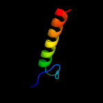
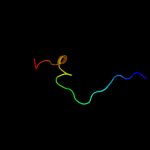
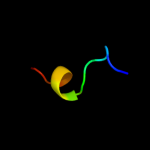
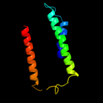
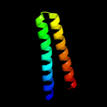
1 d1om2a_
32.8
30
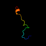
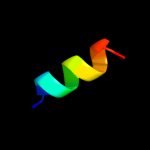
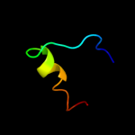
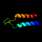
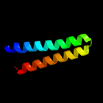
Fold: Open three-helical up-and-down bundleSuperfamily: Mitochondrial import receptor subunit Tom20Family: Mitochondrial import receptor subunit Tom202 d1n1ca_
18.8
17
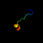
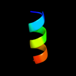
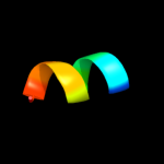
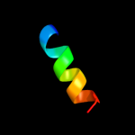
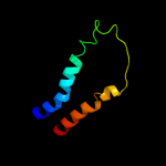
Fold: TorD-likeSuperfamily: TorD-likeFamily: TorD-like3 c2d68A_
14.1
20
PDB header: cell cycleChain: A: PDB Molecule: fop;PDBTitle: structure of the n-terminal domain of fop (fgfr1op) protein
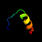
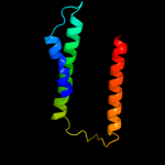
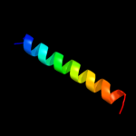
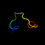
4 c2lc2A_
12.4
33
PDB header: protein bindingChain: A: PDB Molecule: avr3a4;PDBTitle: solution structure of the rxlr effector p. capsici avr3a4
5 c3g43F_
11.8
50
PDB header: metal binding proteinChain: F: PDB Molecule: voltage-dependent l-type calcium channel subunitPDBTitle: crystal structure of the calmodulin-bound cav1.2 c-terminal2 regulatory domain dimer
6 d1p4ea1
10.9
50
Fold: SAM domain-likeSuperfamily: lambda integrase-like, N-terminal domainFamily: lambda integrase-like, N-terminal domain7 c2l0gA_
10.6
36
PDB header: protein bindingChain: A: PDB Molecule: dna polymerase iota;PDBTitle: solution nmr structure of ubiquitin-binding motif (ubm2) of human2 polymerase iota
8 c3b4qA_
10.6
46
PDB header: structural genomics, unknown functionChain: A: PDB Molecule: uncharacterized protein;PDBTitle: crystal structure of a conserved protein domain (unknown2 function) from corynebacterium diphtheriae
9 c2khuA_
10.0
36
PDB header: transferase/protein bindingChain: A: PDB Molecule: immunoglobulin g-binding protein g, dnaPDBTitle: solution structure of the ubiquitin-binding motif of human2 polymerase iota
10 d2ipqx1
10.0
40
Fold: DNA/RNA-binding 3-helical bundleSuperfamily: "Winged helix" DNA-binding domainFamily: STY4665 C-terminal domain-like11 c2axtc_
9.4
21
PDB header: electron transportChain: C: PDB Molecule: photosystem ii cp43 protein;PDBTitle: crystal structure of photosystem ii from thermosynechococcus elongatus
12 d2axtc1
9.4
21
Fold: Photosystem II antenna protein-likeSuperfamily: Photosystem II antenna protein-likeFamily: Photosystem II antenna protein-like13 c3mk7F_
9.2
16
PDB header: oxidoreductaseChain: F: PDB Molecule: cytochrome c oxidase, cbb3-type, subunit p;PDBTitle: the structure of cbb3 cytochrome oxidase
14 d3proc1
8.4
14
Fold: Alpha-lytic protease prodomain-likeSuperfamily: Alpha-lytic protease prodomainFamily: Alpha-lytic protease prodomain15 c2v1sD_
7.7
30
PDB header: oxidoreductaseChain: D: PDB Molecule: mitochondrial import receptor subunit tom20 homolog;PDBTitle: crystal structure of rat tom20-aldh presequence complex
16 c1h2sB_
7.6
14
PDB header: membrane proteinChain: B: PDB Molecule: sensory rhodopsin ii transducer;PDBTitle: molecular basis of transmenbrane signalling by sensory2 rhodopsin ii-transducer complex
17 d1h2sb_
7.6
14
Fold: Transmembrane helix hairpinSuperfamily: Htr2 transmembrane domain-likeFamily: Htr2 transmembrane domain-like18 c1fftG_
7.6
9
PDB header: oxidoreductaseChain: G: PDB Molecule: ubiquinol oxidase;PDBTitle: the structure of ubiquinol oxidase from escherichia coli
19 c2retE_
6.8
25
PDB header: protein transportChain: E: PDB Molecule: pseudopilin epsi;PDBTitle: the crystal structure of a binary complex of two pseudopilins: epsi2 and epsj from the type 2 secretion system of vibrio vulnificus
20 c2qm2B_
6.7
19
PDB header: structural genomics, unknown functionChain: B: PDB Molecule: putative hopj type iii effector protein;PDBTitle: putative hopj type iii effector protein from vibrio parahaemolyticus
21 d2reta1
not modelled
6.3
25
Fold: Pili subunitsSuperfamily: Pili subunitsFamily: GSPII I/J protein-like22 d1c99a_
not modelled
6.3
21
Fold: Transmembrane helix hairpinSuperfamily: F1F0 ATP synthase subunit CFamily: F1F0 ATP synthase subunit C23 d3b2ua2
not modelled
5.8
40
Fold: Knottins (small inhibitors, toxins, lectins)Superfamily: Growth factor receptor domainFamily: Growth factor receptor domain24 d1dxxa1
not modelled
5.7
23
Fold: CH domain-likeSuperfamily: Calponin-homology domain, CH-domainFamily: Calponin-homology domain, CH-domain25 d2fj6a1
not modelled
5.3
25
Fold: SAM domain-likeSuperfamily: YozE-likeFamily: YozE-like26 c3m7kA_
not modelled
5.2
100
PDB header: hydrolase/dnaChain: A: PDB Molecule: restriction endonuclease paci;PDBTitle: crystal structure of paci-dna enzyme product complex
27 d1bhda_
not modelled
5.2
29
Fold: CH domain-likeSuperfamily: Calponin-homology domain, CH-domainFamily: Calponin-homology domain, CH-domain