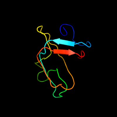
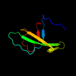
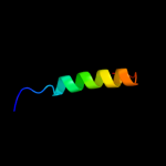
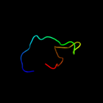
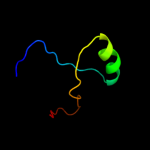
1 c1j6qA_
82.1
24
PDB header: chaperoneChain: A: PDB Molecule: cytochrome c maturation protein e;PDBTitle: solution structure and characterization of the heme2 chaperone ccme
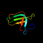
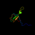
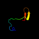
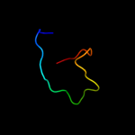
2 d1j6qa_
82.1
24
Fold: OB-foldSuperfamily: Heme chaperone CcmEFamily: Heme chaperone CcmE3 d1sr3a_
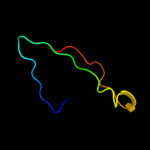
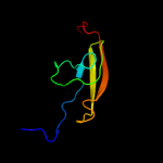
45.5
30
Fold: OB-foldSuperfamily: Heme chaperone CcmEFamily: Heme chaperone CcmE4 c2kctA_
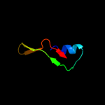
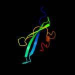
45.1
22
PDB header: chaperoneChain: A: PDB Molecule: cytochrome c-type biogenesis protein ccme;PDBTitle: solution nmr structure of the ob-fold domain of heme2 chaperone ccme from desulfovibrio vulgaris. northeast3 structural genomics target dvr115g.
5 c2r9qD_
39.1
31
PDB header: hydrolaseChain: D: PDB Molecule: 2'-deoxycytidine 5'-triphosphate deaminase;PDBTitle: crystal structure of 2'-deoxycytidine 5'-triphosphate deaminase from2 agrobacterium tumefaciens
6 c2wkdA_
38.6
31
PDB header: dna binding proteinChain: A: PDB Molecule: orf34p2;PDBTitle: crystal structure of a double ile-to-met mutant of protein2 orf34 from lactococcus phage p2
7 c2dzlA_
27.6
16
PDB header: structural genomics unknown functionChain: A: PDB Molecule: protein fam100b;PDBTitle: solution structure of the uba domain in human protein2 fam100b
8 d1n9wa1
27.4
19
Fold: OB-foldSuperfamily: Nucleic acid-binding proteinsFamily: Anticodon-binding domain9 c2wp8J_
25.4
23
PDB header: hydrolaseChain: J: PDB Molecule: exosome complex exonuclease dis3;PDBTitle: yeast rrp44 nuclease
10 c2z14A_
24.6
17
PDB header: signaling proteinChain: A: PDB Molecule: ef-hand domain-containing family member c2;PDBTitle: crystal structure of the n-terminal duf1126 in human ef-2 hand domain containing 2 protein
11 c3h43F_
22.1
23
PDB header: hydrolaseChain: F: PDB Molecule: proteasome-activating nucleotidase;PDBTitle: n-terminal domain of the proteasome-activating nucleotidase2 of methanocaldococcus jannaschii
12 d2ijra1
21.1
21
Fold: Api92-likeSuperfamily: Api92-likeFamily: Api92-like13 c1wydB_
20.6
18
PDB header: ligaseChain: B: PDB Molecule: hypothetical aspartyl-trna synthetase;PDBTitle: crystal structure of aspartyl-trna synthetase from sulfolobus tokodaii
14 d2vnud1
20.3
23
Fold: OB-foldSuperfamily: Nucleic acid-binding proteinsFamily: Cold shock DNA-binding domain-like15 d1t3ta6
19.5
21
Fold: PurM C-terminal domain-likeSuperfamily: PurM C-terminal domain-likeFamily: PurM C-terminal domain-like16 d2pi2e1
19.4
19
Fold: OB-foldSuperfamily: Nucleic acid-binding proteinsFamily: Single strand DNA-binding domain, SSB17 c2wg6L_
19.3
8
PDB header: transcription,hydrolaseChain: L: PDB Molecule: general control protein gcn4,PDBTitle: proteasome-activating nucleotidase (pan) n-domain (57-134)2 from archaeoglobus fulgidus fused to gcn4, p61a mutant
18 c2jwyA_
18.6
15
PDB header: lipoproteinChain: A: PDB Molecule: uncharacterized lipoprotein yaji;PDBTitle: solution nmr structure of uncharacterized lipoprotein yaji from2 escherichia coli. northeast structural genomics target er540
19 d1l0wa1
16.5
25
Fold: OB-foldSuperfamily: Nucleic acid-binding proteinsFamily: Anticodon-binding domain20 c2zauB_
13.2
8
PDB header: transferaseChain: B: PDB Molecule: selenide, water dikinase;PDBTitle: crystal structure of an n-terminally truncated2 selenophosphate synthetase from aquifex aeolicus
21 c1yrlD_
not modelled
12.2
24
PDB header: oxidoreductaseChain: D: PDB Molecule: ketol-acid reductoisomerase;PDBTitle: escherichia coli ketol-acid reductoisomerase
22 c2p39A_
not modelled
11.5
16
PDB header: signaling proteinChain: A: PDB Molecule: fibroblast growth factor 23;PDBTitle: crystal structure of human fgf23
23 c1pwaA_
not modelled
11.4
24
PDB header: hormone/growth factorChain: A: PDB Molecule: fibroblast growth factor-19;PDBTitle: crystal structure of fibroblast growth factor 19
24 d1pwaa_
not modelled
11.4
24
Fold: beta-TrefoilSuperfamily: CytokineFamily: Fibroblast growth factors (FGF)25 c2jn4A_
not modelled
11.2
39
PDB header: structural genomics, unknown functionChain: A: PDB Molecule: hypothetical protein fixu, nift;PDBTitle: solution nmr structure of protein rp4601 from2 rhodopseudomonas palustris. northeast structural genomics3 consortium target rpt2; ontario center for structural4 proteomics target rp4601.
26 d2jn4a1
not modelled
11.2
39
Fold: NifT/FixU barrel-likeSuperfamily: NifT/FixU-likeFamily: NifT/FixU27 c2zodB_
not modelled
11.1
8
PDB header: transferaseChain: B: PDB Molecule: selenide, water dikinase;PDBTitle: crystal structure of selenophosphate synthetase from2 aquifex aeolicus
28 c2pqaB_
not modelled
10.9
10
PDB header: replicationChain: B: PDB Molecule: replication protein a 14 kda subunit;PDBTitle: crystal structure of full-length human rpa 14/32 heterodimer
29 d1so0a_
not modelled
10.6
23
Fold: SupersandwichSuperfamily: Galactose mutarotase-likeFamily: Aldose 1-epimerase (mutarotase)30 c1b8aB_
not modelled
10.4
24
PDB header: ligaseChain: B: PDB Molecule: protein (aspartyl-trna synthetase);PDBTitle: aspartyl-trna synthetase
31 c1vqwB_
not modelled
8.7
16
PDB header: structural genomics, unknown functionChain: B: PDB Molecule: protein with similarity to flavin-containingPDBTitle: crystal structure of a protein with similarity to flavin-2 containing monooxygenases and to mammalian dimethylalanine3 monooxygenases
32 d1ps9a2
not modelled
8.5
15
Fold: FAD/NAD(P)-binding domainSuperfamily: FAD/NAD(P)-binding domainFamily: C-terminal domain of adrenodoxin reductase-like33 c2pi2A_
not modelled
7.1
11
PDB header: replication, dna binding proteinChain: A: PDB Molecule: replication protein a 32 kda subunit;PDBTitle: full-length replication protein a subunits rpa14 and rpa32
34 d2zoda2
not modelled
6.9
5
Fold: PurM C-terminal domain-likeSuperfamily: PurM C-terminal domain-likeFamily: PurM C-terminal domain-like35 d1c0aa1
not modelled
6.2
18
Fold: OB-foldSuperfamily: Nucleic acid-binding proteinsFamily: Anticodon-binding domain36 d1b33n_
not modelled
6.1
63
Fold: Allophycocyanin linker chain (domain)Superfamily: Allophycocyanin linker chain (domain)Family: Allophycocyanin linker chain (domain)37 d2z1ea2
not modelled
6.1
17
Fold: PurM C-terminal domain-likeSuperfamily: PurM C-terminal domain-likeFamily: PurM C-terminal domain-like38 c3ls1A_
not modelled
6.0
16
PDB header: photosynthesisChain: A: PDB Molecule: sll1638 protein;PDBTitle: crystal structure of cyanobacterial psbq from synechocystis2 sp. pcc 6803 complexed with zn2+
39 d1yloa1
not modelled
5.7
13
Fold: Domain of alpha and beta subunits of F1 ATP synthase-likeSuperfamily: Aminopeptidase/glucanase lid domainFamily: Aminopeptidase/glucanase lid domain40 d1krta_
not modelled
5.6
6
Fold: OB-foldSuperfamily: Nucleic acid-binding proteinsFamily: Anticodon-binding domain41 d1geha2
not modelled
5.6
8
Fold: Ferredoxin-likeSuperfamily: RuBisCO, large subunit, small (N-terminal) domainFamily: Ribulose 1,5-bisphosphate carboxylase-oxygenase42 d2gycb1
not modelled
5.4
25
Fold: Reductase/isomerase/elongation factor common domainSuperfamily: Translation proteinsFamily: Ribosomal protein L343 c2esyA_
not modelled
5.2
36
PDB header: lipid binding proteinChain: A: PDB Molecule: lung surfactant protein c;PDBTitle: structure and influence on stability and activity of the n-2 terminal propetide part of lung surfactant protein c
44 c3m9bK_
not modelled
5.1
20
PDB header: chaperoneChain: K: PDB Molecule: proteasome-associated atpase;PDBTitle: crystal structure of the amino terminal coiled coil domain and the2 inter domain of the mycobacterium tuberculosis proteasomal atpase mpa
45 c2pxgA_
not modelled
5.1
14
PDB header: membrane proteinChain: A: PDB Molecule: outer membrane protein;PDBTitle: nmr solution structure of omla
46 d1lura_
not modelled
5.0
18
Fold: SupersandwichSuperfamily: Galactose mutarotase-likeFamily: Aldose 1-epimerase (mutarotase)