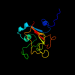
| 1 |
|
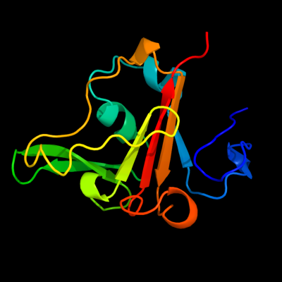
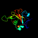
PDB 3fhh chain A
Region: 39 - 167
Aligned: 126
Modelled: 129
Confidence: 100.0%
Identity: 33%
PDB header:membrane protein
Chain: A: PDB Molecule:outer membrane heme receptor shua;
PDBTitle: crystal structure of the heme/hemoglobin outer membrane2 transporter shua from shigella dysenteriae
Phyre2
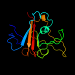
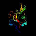
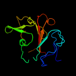
| 2 |
|
PDB 2hdi chain A
Region: 39 - 167
Aligned: 128
Modelled: 129
Confidence: 100.0%
Identity: 45%
PDB header:protein transport,antimicrobial protein
Chain: A: PDB Molecule:colicin i receptor;
PDBTitle: crystal structure of the colicin i receptor cir from e.coli in complex2 with receptor binding domain of colicin ia.
Phyre2
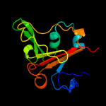
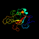
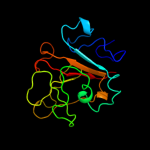
| 3 |
|
PDB 3qlb chain A
Region: 29 - 167
Aligned: 132
Modelled: 139
Confidence: 100.0%
Identity: 27%
PDB header:metal transport
Chain: A: PDB Molecule:enantio-pyochelin receptor;
PDBTitle: enantiopyochelin outer membrane tonb-dependent transporter from2 pseudomonas fluorescens bound to the ferri-enantiopyochelin
Phyre2
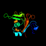
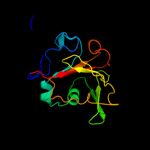
| 4 |
|
PDB 3efm chain A
Region: 26 - 167
Aligned: 137
Modelled: 142
Confidence: 100.0%
Identity: 18%
PDB header:membrane protein
Chain: A: PDB Molecule:ferric alcaligin siderophore receptor;
PDBTitle: structure of the alcaligin outer membrane recepteur faua from2 bordetella pertussis
Phyre2
| 5 |
|
PDB 1xkw chain A
Region: 32 - 167
Aligned: 128
Modelled: 136
Confidence: 100.0%
Identity: 23%
PDB header:membrane protein
Chain: A: PDB Molecule:fe(iii)-pyochelin receptor;
PDBTitle: pyochelin outer membrane receptor fpta from pseudomonas2 aeruginosa
Phyre2
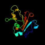
| 6 |
|
PDB 1by5 chain A
Region: 27 - 167
Aligned: 136
Modelled: 141
Confidence: 100.0%
Identity: 25%
Fold: Transmembrane beta-barrels
Superfamily: Porins
Family: Ligand-gated protein channel
Phyre2
| 7 |
|
PDB 2guf chain A domain 1
Region: 39 - 167
Aligned: 123
Modelled: 129
Confidence: 100.0%
Identity: 38%
Fold: Transmembrane beta-barrels
Superfamily: Porins
Family: Ligand-gated protein channel
Phyre2
| 8 |
|
PDB 1kmo chain A
Region: 40 - 167
Aligned: 128
Modelled: 128
Confidence: 100.0%
Identity: 27%
Fold: Transmembrane beta-barrels
Superfamily: Porins
Family: Ligand-gated protein channel
Phyre2
| 9 |
|
PDB 1xkh chain C
Region: 21 - 167
Aligned: 143
Modelled: 139
Confidence: 100.0%
Identity: 27%
PDB header:membrane protein
Chain: C: PDB Molecule:ferripyoverdine receptor;
PDBTitle: pyoverdine outer membrane receptor fpva from pseudomonas aeruginosa2 pao1 bound to pyoverdine
Phyre2
| 10 |
|
PDB 2grx chain B
Region: 28 - 167
Aligned: 135
Modelled: 140
Confidence: 100.0%
Identity: 25%
PDB header:metal transport
Chain: B: PDB Molecule:ferrichrome-iron receptor;
PDBTitle: crystal structure of tonb in complex with fhua, e. coli2 outer membrane receptor for ferrichrome
Phyre2
| 11 |
|
PDB 2iah chain A
Region: 26 - 167
Aligned: 138
Modelled: 142
Confidence: 100.0%
Identity: 27%
PDB header:membrane protein
Chain: A: PDB Molecule:ferripyoverdine receptor;
PDBTitle: crystal structure of the ferripyoverdine receptor of the outer2 membrane of pseudomonas aeruginosa bound to ferripyoverdine.
Phyre2
| 12 |
|
PDB 1po3 chain A
Region: 50 - 167
Aligned: 118
Modelled: 118
Confidence: 100.0%
Identity: 28%
PDB header:membrane protein
Chain: A: PDB Molecule:iron(iii) dicitrate transport protein feca
PDBTitle: crystal structure of ferric citrate transporter feca in2 complex with ferric citrate
Phyre2
| 13 |
|
PDB 3csl chain B
Region: 49 - 167
Aligned: 119
Modelled: 119
Confidence: 100.0%
Identity: 24%
PDB header:membrane protein/heme binding protein
Chain: B: PDB Molecule:hasr protein;
PDBTitle: structure of the serratia marcescens hemophore receptor hasr in2 complex with its hemophore hasa and heme
Phyre2