Phyre Home Retrieve Phyre Job Id
Emaill.a.kelley@imperial.ac.uk
DescriptionP62723
DateThu Jan 5 12:07:48 GMT 2012
Unique Job ID3c783e027de8fce4

Summary 

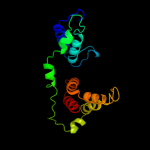
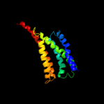
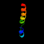
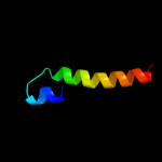
Top model
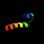
Image coloured by rainbow N → C terminus
Model (left) based on template c1zcdA_
Top template information
PDB header:membrane protein
Chain: A: PDB Molecule:na(+)/h(+) antiporter 1;
PDBTitle: crystal structure of the na+/h+ antiporter nhaa
Confidence and coverage
Confidence: 60.9% Coverage: 46%
159 residues ( 46% of your sequence) have been modelled with 60.9% confidence by the single highest scoring template.
You may wish to submit your sequence to Phyrealarm. This will automatically scan your sequence every week for new potential templates as they appear in the Phyre2 library.
Please note: You must be registered and logged in to use Phyrealarm.
3D viewing
Interactive 3D view in Jmol

Sequence analysis 

Secondary structure and disorder prediction 

   1........10.........20.........30.........40.........50.........60
Sequence  MTNITLQKQHRTLWHFIPGLALSAVITGVALWGGSIPAVAGAGFSALTLAILLGMVLGNT
Secondary structure 


SS confidence 


Disorder  ???


??


?


Disorder confidence 


   .........70.........80.........90.........100.........110.........120
Sequence  IYPHIWKSCDGGVLFAKQYLLRLGIILYGFRLTFSQIADVGISGIIIDVLTLSSTFLLAC
Secondary structure 


SS confidence 


Disorder 


???


Disorder confidence 


   .........130.........140.........150.........160.........170.........180
Sequence  FLGQKVFGLDKHTSWLIGAGSSICGAAAVLATEPVVKAEASKVTVAVATVVIFGTVAIFL
Secondary structure 


SS confidence 


Disorder 


??


??
?

?


Disorder confidence 


   .........190.........200.........210.........220.........230.........240
Sequence  YPAIYPLMSQWFSPETFGIYIGSTVHEVAQVVAAGHAISPDAENAAVISKMLRVMMLAPF
Secondary structure 


SS confidence 


Disorder 


?


Disorder confidence 


   .........250.........260.........270.........280.........290.........300
Sequence  LILLAARVKQLSGANSGEKSKITIPWFAILFIVVAIFNSFHLLPQSVVNMLVTLDTFLLA
Secondary structure 


SS confidence 


Disorder 


????????????


Disorder confidence 


   .........310.........320.........330.........340.........
Sequence  MAMAALGLTTHVSALKKAGAKPLLMALVLFAWLIVGGGAINYVIQSVIA
Secondary structure 


SS confidence 


Disorder 


?


?


??
Disorder confidence 


Confidence Key
High(9)                    Low (0)
?Disordered
Alpha helix
Beta strand

Domain analysis 

Hover over an aligned region to see model and summary info

Please note, only up to the top 20 hits are modelled to reduce computer load

RankAligned region

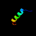
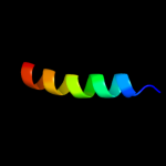
PDB 1zcd chain A

3D model

Region: 9 - 176
Aligned: 159
Modelled: 168
Confidence: 60.9%
Identity: 12%
PDB header:membrane protein
Chain: A: PDB Molecule:na(+)/h(+) antiporter 1;
PDBTitle: crystal structure of the na+/h+ antiporter nhaa

Phyre2

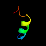
PDB 3lr4 chain A

3D model

Region: 220 - 237
Aligned: 18
Modelled: 18
Confidence: 34.1%
Identity: 28%
PDB header:transferase
Chain: A: PDB Molecule:sensor protein;
PDBTitle: periplasmic domain of the riss sensor protein from burkholderia2 pseudomallei, barium phased at low ph

Phyre2

PDB 2yvx chain D

3D model

Region: 261 - 348
Aligned: 88
Modelled: 88
Confidence: 24.3%
Identity: 11%
PDB header:transport protein
Chain: D: PDB Molecule:mg2+ transporter mgte;
PDBTitle: crystal structure of magnesium transporter mgte

Phyre2

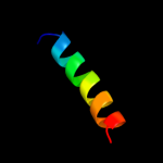
PDB 1woz chain A

3D model

Region: 273 - 302
Aligned: 30
Modelled: 28
Confidence: 21.3%
Identity: 20%
PDB header:structural genomics, unknown function
Chain: A: PDB Molecule:177aa long conserved hypothetical protein (st1454);
PDBTitle: crystal structure of uncharacterized protein st1454 from sulfolobus2 tokodaii

Phyre2

PDB 2gke chain A domain 2

3D model

Region: 137 - 159
Aligned: 23
Modelled: 23
Confidence: 19.2%
Identity: 26%
Fold: Diaminopimelate epimerase-like
Superfamily: Diaminopimelate epimerase-like
Family: Diaminopimelate epimerase

Phyre2

PDB 1pw4 chain A

3D model

Region: 98 - 265
Aligned: 161
Modelled: 168
Confidence: 15.9%
Identity: 11%
Fold: MFS general substrate transporter
Superfamily: MFS general substrate transporter
Family: Glycerol-3-phosphate transporter

Phyre2

PDB 1e3h chain A domain 2

3D model

Region: 142 - 155
Aligned: 14
Modelled: 14
Confidence: 14.4%
Identity: 21%
Fold: Ribosomal protein S5 domain 2-like
Superfamily: Ribosomal protein S5 domain 2-like
Family: Ribonuclease PH domain 1-like

Phyre2

PDB 3efe chain C

3D model

Region: 142 - 160
Aligned: 19
Modelled: 19
Confidence: 13.8%
Identity: 32%
PDB header:chaperone
Chain: C: PDB Molecule:thij/pfpi family protein;
PDBTitle: the crystal structure of the thij/pfpi family protein from bacillus2 anthracis

Phyre2

PDB 2idx chain A

3D model

Region: 283 - 302
Aligned: 20
Modelled: 20
Confidence: 13.1%
Identity: 25%
PDB header:transferase
Chain: A: PDB Molecule:cob(i)yrinic acid a,c-diamide
PDBTitle: structure of human atp:cobalamin adenosyltransferase bound2 to atp.

Phyre2

PDB 1wy1 chain B

3D model

Region: 274 - 302
Aligned: 29
Modelled: 29
Confidence: 12.8%
Identity: 21%
PDB header:transferase
Chain: B: PDB Molecule:hypothetical protein ph0671;
PDBTitle: crystal structure of the ph0671 protein from pyrococcus horikoshii ot3

Phyre2

PDB 1nog chain A

3D model

Region: 274 - 302
Aligned: 29
Modelled: 29
Confidence: 12.1%
Identity: 14%
Fold: Ferritin-like
Superfamily: Cobalamin adenosyltransferase-like
Family: Cobalamin adenosyltransferase

Phyre2

PDB 1nog chain A

3D model

Region: 274 - 302
Aligned: 29
Modelled: 29
Confidence: 12.1%
Identity: 14%
PDB header:structural genomics, unknown function
Chain: A: PDB Molecule:conserved hypothetical protein ta0546;
PDBTitle: crystal structure of conserved protein 0546 from thermoplasma2 acidophilum

Phyre2

PDB 1zq1 chain C domain 1

3D model

Region: 312 - 333
Aligned: 22
Modelled: 22
Confidence: 12.0%
Identity: 36%
Fold: GatB/YqeY motif
Superfamily: GatB/YqeY motif
Family: GatB/GatE C-terminal domain-like

Phyre2

PDB 2ah6 chain B

3D model

Region: 274 - 302
Aligned: 29
Modelled: 29
Confidence: 11.3%
Identity: 21%
PDB header:transferase
Chain: B: PDB Molecule:bh1595, unknown conserved protein;
PDBTitle: crystal structure of a putative cobalamin adenosyltransferase (bh1595)2 from bacillus halodurans c-125 at 1.60 a resolution

Phyre2

PDB 2g2d chain A

3D model

Region: 283 - 302
Aligned: 20
Modelled: 20
Confidence: 11.3%
Identity: 15%
PDB header:transferase
Chain: A: PDB Molecule:atp:cobalamin adenosyltransferase;
PDBTitle: crystal structure of a putative pduo-type atp:cobalamin2 adenosyltransferase from mycobacterium tuberculosis

Phyre2

PDB 1rty chain A

3D model

Region: 274 - 302
Aligned: 29
Modelled: 29
Confidence: 11.1%
Identity: 21%
Fold: Ferritin-like
Superfamily: Cobalamin adenosyltransferase-like
Family: Cobalamin adenosyltransferase

Phyre2

PDB 3f5d chain A

3D model

Region: 142 - 160
Aligned: 19
Modelled: 19
Confidence: 10.4%
Identity: 32%
PDB header:structural genomics, unknown function
Chain: A: PDB Molecule:protein ydea;
PDBTitle: crystal structure of a protein of unknown function from2 bacillus subtilis

Phyre2

PDB 2nt8 chain A

3D model

Region: 274 - 305
Aligned: 32
Modelled: 32
Confidence: 10.3%
Identity: 19%
PDB header:transferase
Chain: A: PDB Molecule:cobalamin adenosyltransferase;
PDBTitle: atp bound at the active site of a pduo type atp:co(i)rrinoid2 adenosyltransferase from lactobacillus reuteri

Phyre2

PDB 2zhz chain C

3D model

Region: 274 - 302
Aligned: 29
Modelled: 29
Confidence: 10.2%
Identity: 14%
PDB header:transferase
Chain: C: PDB Molecule:atp:cob(i)alamin adenosyltransferase, putative;
PDBTitle: crystal structure of a pduo-type atp:cobalamin adenosyltransferase2 from burkholderia thailandensis

Phyre2

PDB 3ke4 chain B

3D model

Region: 274 - 305
Aligned: 32
Modelled: 32
Confidence: 10.0%
Identity: 16%
PDB header:transferase
Chain: B: PDB Molecule:hypothetical cytosolic protein;
PDBTitle: crystal structure of a pduo-type atp:cob(i)alamin adenosyltransferase2 from bacillus cereus

Phyre2
1

c1zcdA_
2

c3lr4A_
3

c2yvxD_
4

c1wozA_
5

d2gkea2
6

d1pw4a_
7

d1e3ha2
8

c3efeC_
9

c2idxA_
10

c1wy1B_
11

d1noga_
12

c1nogA_
13

d1zq1c1
14

c2ah6B_
15

c2g2dA_
16

d1rtya_
17

c3f5dA_
18

c2nt8A_
19

c2zhzC_
20

c3ke4B_
21


22


23


24


25


26


27


28


29


30


31


32


33


34


35


36


37


38


Detailed template information 

#
Template Alignment Coverage3D Model Confidence
% i.d. Template Information
1c1zcdA_


60.9 12 PDB header:membrane protein
Chain: A: PDB Molecule:na(+)/h(+) antiporter 1;
PDBTitle: crystal structure of the na+/h+ antiporter nhaa
2c3lr4A_


34.1 28 PDB header:transferase
Chain: A: PDB Molecule:sensor protein;
PDBTitle: periplasmic domain of the riss sensor protein from burkholderia2 pseudomallei, barium phased at low ph
3c2yvxD_


24.3 11 PDB header:transport protein
Chain: D: PDB Molecule:mg2+ transporter mgte;
PDBTitle: crystal structure of magnesium transporter mgte
4c1wozA_


21.3 20 PDB header:structural genomics, unknown function
Chain: A: PDB Molecule:177aa long conserved hypothetical protein (st1454);
PDBTitle: crystal structure of uncharacterized protein st1454 from sulfolobus2 tokodaii
5d2gkea2


19.2 26 Fold:Diaminopimelate epimerase-like
Superfamily:Diaminopimelate epimerase-like
Family:Diaminopimelate epimerase
6d1pw4a_


15.9 11 Fold:MFS general substrate transporter
Superfamily:MFS general substrate transporter
Family:Glycerol-3-phosphate transporter
7d1e3ha2


14.4 21 Fold:Ribosomal protein S5 domain 2-like
Superfamily:Ribosomal protein S5 domain 2-like
Family:Ribonuclease PH domain 1-like
8c3efeC_


13.8 32 PDB header:chaperone
Chain: C: PDB Molecule:thij/pfpi family protein;
PDBTitle: the crystal structure of the thij/pfpi family protein from bacillus2 anthracis
9c2idxA_


13.1 25 PDB header:transferase
Chain: A: PDB Molecule:cob(i)yrinic acid a,c-diamide
PDBTitle: structure of human atp:cobalamin adenosyltransferase bound2 to atp.
10c1wy1B_


12.8 21 PDB header:transferase
Chain: B: PDB Molecule:hypothetical protein ph0671;
PDBTitle: crystal structure of the ph0671 protein from pyrococcus horikoshii ot3
11d1noga_


12.1 14 Fold:Ferritin-like
Superfamily:Cobalamin adenosyltransferase-like
Family:Cobalamin adenosyltransferase
12c1nogA_


12.1 14 PDB header:structural genomics, unknown function
Chain: A: PDB Molecule:conserved hypothetical protein ta0546;
PDBTitle: crystal structure of conserved protein 0546 from thermoplasma2 acidophilum
13d1zq1c1


12.0 36 Fold:GatB/YqeY motif
Superfamily:GatB/YqeY motif
Family:GatB/GatE C-terminal domain-like
14c2ah6B_


11.3 21 PDB header:transferase
Chain: B: PDB Molecule:bh1595, unknown conserved protein;
PDBTitle: crystal structure of a putative cobalamin adenosyltransferase (bh1595)2 from bacillus halodurans c-125 at 1.60 a resolution
15c2g2dA_


11.3 15 PDB header:transferase
Chain: A: PDB Molecule:atp:cobalamin adenosyltransferase;
PDBTitle: crystal structure of a putative pduo-type atp:cobalamin2 adenosyltransferase from mycobacterium tuberculosis
16d1rtya_


11.1 21 Fold:Ferritin-like
Superfamily:Cobalamin adenosyltransferase-like
Family:Cobalamin adenosyltransferase
17c3f5dA_


10.4 32 PDB header:structural genomics, unknown function
Chain: A: PDB Molecule:protein ydea;
PDBTitle: crystal structure of a protein of unknown function from2 bacillus subtilis
18c2nt8A_


10.3 19 PDB header:transferase
Chain: A: PDB Molecule:cobalamin adenosyltransferase;
PDBTitle: atp bound at the active site of a pduo type atp:co(i)rrinoid2 adenosyltransferase from lactobacillus reuteri
19c2zhzC_


10.2 14 PDB header:transferase
Chain: C: PDB Molecule:atp:cob(i)alamin adenosyltransferase, putative;
PDBTitle: crystal structure of a pduo-type atp:cobalamin adenosyltransferase2 from burkholderia thailandensis
20c3ke4B_


10.0 16 PDB header:transferase
Chain: B: PDB Molecule:hypothetical cytosolic protein;
PDBTitle: crystal structure of a pduo-type atp:cob(i)alamin adenosyltransferase2 from bacillus cereus
21d2fexa1


not modelled 9.9 37 Fold:Flavodoxin-like
Superfamily:Class I glutamine amidotransferase-like
Family:DJ-1/PfpI
22c3ci1A_


not modelled 9.5 19 PDB header:transferase
Chain: A: PDB Molecule:cobalamin adenosyltransferase pduo-like protein;
PDBTitle: structure of the pduo-type atp:co(i)rrinoid2 adenosyltransferase from lactobacillus reuteri complexed3 with four-coordinate cob(ii)alamin and atp
23d1fftb2


not modelled 9.0 22 Fold:Transmembrane helix hairpin
Superfamily:Cytochrome c oxidase subunit II-like, transmembrane region
Family:Cytochrome c oxidase subunit II-like, transmembrane region
24c2hp0A_


not modelled 9.0 21 PDB header:isomerase
Chain: A: PDB Molecule:ids-epimerase;
PDBTitle: crystal structure of iminodisuccinate epimerase
25d1rtyb_


not modelled 8.8 21 Fold:Ferritin-like
Superfamily:Cobalamin adenosyltransferase-like
Family:Cobalamin adenosyltransferase
26c1wvtA_


not modelled 7.9 16 PDB header:structural genomics, unknown function
Chain: A: PDB Molecule:hypothetical protein st2180;
PDBTitle: crystal structure of uncharacterized protein st2180 from sulfolobus2 tokodaii
27d1z96a1


not modelled 7.8 35 Fold:RuvA C-terminal domain-like
Superfamily:UBA-like
Family:UBA domain
28c2g38A_


not modelled 7.5 23 PDB header:structural genomics, unknown function
Chain: A: PDB Molecule:pe family protein;
PDBTitle: a pe/ppe protein complex from mycobacterium tuberculosis
29d2g38a1


not modelled 7.5 23 Fold:Ferritin-like
Superfamily:PE/PPE dimer-like
Family:PE
30d1rfza_


not modelled 7.3 14 Fold:YutG-like
Superfamily:YutG-like
Family:YutG-like
31d1tlqa_


not modelled 6.7 7 Fold:YutG-like
Superfamily:YutG-like
Family:YutG-like
32c1tlqA_


not modelled 6.7 7 PDB header:structural genomics, unknown function
Chain: A: PDB Molecule:hypothetical protein ypjq;
PDBTitle: crystal structure of protein ypjq from bacillus subtilis, pfam duf64
33d1szqa_


not modelled 6.5 9 Fold:2-methylcitrate dehydratase PrpD
Superfamily:2-methylcitrate dehydratase PrpD
Family:2-methylcitrate dehydratase PrpD
34c3mgkA_


not modelled 6.4 21 PDB header:structural genomics, unknown function
Chain: A: PDB Molecule:intracellular protease/amidase related enzyme
PDBTitle: crystal structure of probable protease/amidase from2 clostridium acetobutylicum atcc 824
35c6rlxB_


not modelled 6.1 42 PDB header:hormone(muscle relaxant)
Chain: B: PDB Molecule:relaxin, b-chain;
PDBTitle: x-ray structure of human relaxin at 1.5 angstroms. comparison to2 insulin and implications for receptor binding determinants
36d1vf5b_


not modelled 6.0 13 Fold:a domain/subunit of cytochrome bc1 complex (Ubiquinol-cytochrome c reductase)
Superfamily:a domain/subunit of cytochrome bc1 complex (Ubiquinol-cytochrome c reductase)
Family:a domain/subunit of cytochrome bc1 complex (Ubiquinol-cytochrome c reductase)
37d3dtub2


not modelled 5.9 16 Fold:Transmembrane helix hairpin
Superfamily:Cytochrome c oxidase subunit II-like, transmembrane region
Family:Cytochrome c oxidase subunit II-like, transmembrane region
38d1sy7a1


not modelled 5.1 11 Fold:Flavodoxin-like
Superfamily:Class I glutamine amidotransferase-like
Family:Catalase, C-terminal domain

Binding site prediction 

Due to computational demand, binding site predictions are not run for batch jobs

If you want to predict binding sites, please manually submit your model of choice to 3DLigandSite


Phyre is for academic use only

Please cite: Protein structure prediction on the web: a case study using the Phyre server
Kelley LA and Sternberg MJE. Nature Protocols 4, 363 - 371 (2009) [pdf] [Import into BibTeX]
 
If you use the binding site predictions from 3DLigandSite, please also cite:
3DLigandSite: predicting ligand-binding sites using similar structures.
Wass MN, Kelley LA and Sternberg MJ Nucleic Acids Research 38, W469-73 (2010) [PubMed]
 
© Structural Bioinformatics Group
Imperial College London
Lawrence Kelley, Benjamin Jefferys
Disclaimer
Terms and Conditions
Component software
Template detection: HHpred 1.51
Secondary structure prediction: Psi-pred 2.5
Disorder prediction: Disopred 2.4
Transmembrane prediction: Memsat_SVM
Multi-template modelling and ab initio: Poing 1.0