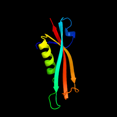
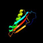
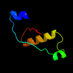
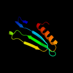
PDB header:membrane protein Chain: A: PDB Molecule:putative outer membrane protein, signal; PDBTitle: crystal structure of the e. coli outer membrane lipoprotein2 rcsf
Confidence and coverage
Confidence:
100.0%
Coverage:
64%
86 residues ( 64% of your sequence) have been modelled with 100.0% confidence by the single highest scoring template.
You may wish to submit your sequence to Phyrealarm. This will automatically scan your sequence every week for new potential templates as they appear in the Phyre2 library.
Region: 59 - 131 Aligned: 73 Modelled: 73 Confidence: 96.8% Identity: 10% PDB header:structural genomics, unknown function Chain: B: PDB Molecule:uncharacterized protein; PDBTitle: crystal structure of a protein with unknown function which belongs to2 pfam duf74 family (pepe_0654) from pediococcus pentosaceus atcc 257453 at 2.73 a resolution
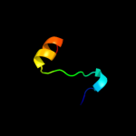
Region: 86 - 103 Aligned: 18 Modelled: 18 Confidence: 12.0% Identity: 28% PDB header:protein binding Chain: B: PDB Molecule:peptide of far upstream element-binding protein 1; PDBTitle: solution structure of the first two rrm domains of fir in the complex2 with fbp nbox peptide
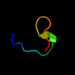
Region: 105 - 124 Aligned: 17 Modelled: 17 Confidence: 5.6% Identity: 29% PDB header:proteinase inhibitor (trypsin) Chain: A: PDB Molecule:trypsin inhibitor; PDBTitle: determination of the complete three-dimensional structure2 of the trypsin inhibitor from squash seeds in aqueous3 solution by nuclear magnetic resonance and a combination4 of distance geometry and dynamical simulated annealing
Phyre2
21
22
23
24
25
Detailed template information
Binding site prediction
Due to computational demand, binding site predictions are not run for batch jobs
If you want to predict binding sites, please manually submit your model of choice to 3DLigandSite
Transmembrane helix prediction
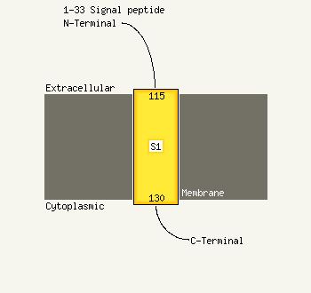
Transmembrane helices have been predicted in your sequence to adopt the topology shown below
Phyre is for academic use only
Please cite: Protein structure prediction on
the web: a case study using the Phyre server
Kelley LA and Sternberg MJE. Nature Protocols
4, 363 - 371 (2009) [pdf] [Import into BibTeX]
If you use the binding site
predictions from 3DLigandSite, please also cite:
3DLigandSite: predicting ligand-binding sites using similar structures.
Wass MN, Kelley LA and Sternberg
MJ Nucleic Acids Research 38, W469-73 (2010) [PubMed]