| Secondary structure and disorder prediction | |
| | |
1 | . | . | . | . | . | . | . | . | 10 | . | . | . | . | . | . | . | . | . | 20 | . | . | . | . | . | . | . | . | . | 30 | . | . |
| Sequence | |
M | T | T | S | M | L | N | A | K | L | L | P | T | A | P | S | A | A | V | V | V | V | R | V | V | V | V | V | G | N | A | P |
| Secondary structure | |
|
|
|  | | | | | | | |  |
|
|
|
|
| | | | | | | | | | | | |
|
|
|
| SS confidence | |
|
|
|
|
|
|
|
|
|
|
|
|
|
|
|
|
|
|
|
|
|
|
|
|
|
|
|
|
|
|
|
|
| Disorder | |
? | ? | ? | ? | ? | ? | ? | ? |
|
| ? | ? | ? |
|
|
|
|
|
|
|
|
|
|
|
|
| ? | ? | ? | ? | ? | ? |
| Disorder confidence | |
|
|
|
|
|
|
|
|
|
|
|
|
|
|
|
|
|
|
|
|
|
|
|
|
|
|
|
|
|
|
|
|
| |
| Confidence Key |
| High(9) | |
|
|
|
|
|
|
|
|
|
Low (0) |
| ? | Disordered |
 | Alpha helix |
| Beta strand |
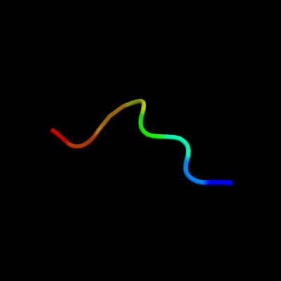
Hover over an aligned region to see model and summary info
Please note, only up to the top 20 hits are modelled to reduce computer load
|
| 1 |
|
PDB 1v9c chain A
Region: 25 - 32
Aligned: 8
Modelled: 8
Confidence: 16.8%
Identity: 75%
Fold: Flavodoxin-like
Superfamily: Precorrin-8X methylmutase CbiC/CobH
Family: Precorrin-8X methylmutase CbiC/CobH
Phyre2
| 2 |
|
PDB 1f2v chain A
Region: 25 - 32
Aligned: 8
Modelled: 8
Confidence: 13.1%
Identity: 75%
Fold: Flavodoxin-like
Superfamily: Precorrin-8X methylmutase CbiC/CobH
Family: Precorrin-8X methylmutase CbiC/CobH
Phyre2
| 3 |
|
PDB 2afv chain B
Region: 25 - 32
Aligned: 8
Modelled: 8
Confidence: 12.5%
Identity: 63%
PDB header:isomerase
Chain: B: PDB Molecule:cobalamin biosynthesis precorrin isomerase;
PDBTitle: the crystal structure of putative precorrin isomerase cbic2 in cobalamin biosynthesis
Phyre2
| 4 |
|
PDB 3e7d chain C
Region: 25 - 32
Aligned: 8
Modelled: 8
Confidence: 11.9%
Identity: 63%
PDB header:isomerase
Chain: C: PDB Molecule:cobh, precorrin-8x methylmutase;
PDBTitle: crystal structure of precorrin-8x methyl mutase cbic/cobh from2 brucella melitensis
Phyre2
| 5 |
|
PDB 1ou0 chain A
Region: 25 - 32
Aligned: 8
Modelled: 8
Confidence: 11.8%
Identity: 75%
Fold: Flavodoxin-like
Superfamily: Precorrin-8X methylmutase CbiC/CobH
Family: Precorrin-8X methylmutase CbiC/CobH
Phyre2
|
| Detailed template information | |
Due to computational demand, binding site predictions are not run for batch jobs
If you want to predict binding sites, please manually submit your model of choice to 3DLigandSite
Phyre is for academic use only
| Please cite: Protein structure prediction on
the web: a case study using the Phyre server |
| Kelley LA and Sternberg MJE. Nature Protocols
4, 363 - 371 (2009) [pdf] [Import into BibTeX] |
| |
| If you use the binding site
predictions from 3DLigandSite, please also cite: |
| 3DLigandSite: predicting ligand-binding sites using similar structures. |
| Wass MN, Kelley LA and Sternberg
MJ Nucleic Acids Research 38, W469-73 (2010) [PubMed] |
| |
|
|
|
|