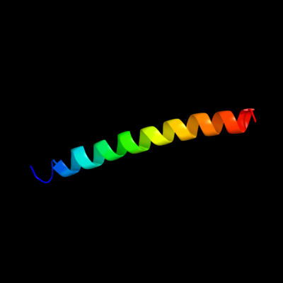
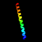
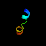
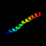
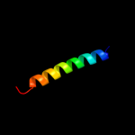
PDB header:blood clotting Chain: A: PDB Molecule:fibrin alpha-1 chain; PDBTitle: fibrin d-dimer, lamprey complexed with the peptide ligand: gly-his-2 arg-pro-amide
Confidence and coverage
Confidence:
66.6%
Coverage:
22%
34 residues ( 22% of your sequence) have been modelled with 66.6% confidence by the single highest scoring template.
You may wish to submit your sequence to Phyrealarm. This will automatically scan your sequence every week for new potential templates as they appear in the Phyre2 library.
Please note: You must be registered and logged in to use Phyrealarm.
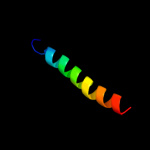
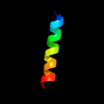
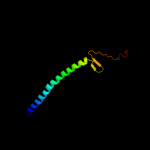
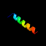
Region: 1 - 21 Aligned: 21 Modelled: 21 Confidence: 13.1% Identity: 19% PDB header:membrane protein, protein transport Chain: E: PDB Molecule:preprotein translocase subunit secg; PDBTitle: crystal structure of the protein-translocation complex formed by the2 secy channel and the seca atpase
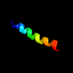
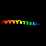
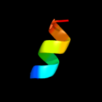
Region: 27 - 111 Aligned: 73 Modelled: 85 Confidence: 8.2% Identity: 10% PDB header:contractile protein Chain: C: PDB Molecule:microtubule motor protein ncd; PDBTitle: the crystal structure of a minus-end directed microtubule2 motor protein ncd reveals variable dimer conformations