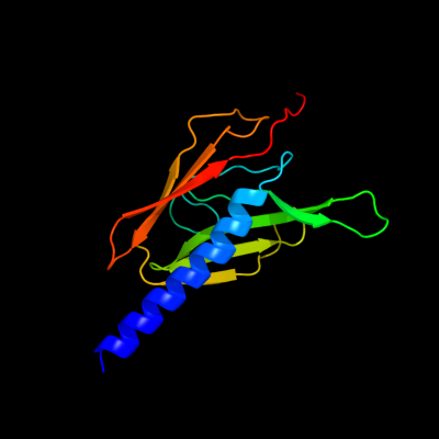
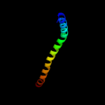
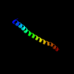
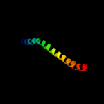
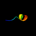
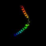
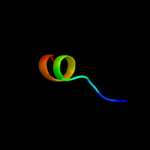
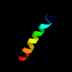
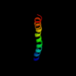
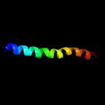
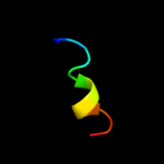
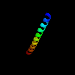
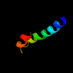
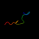
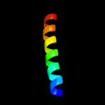
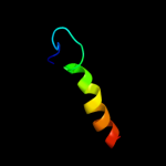
Phyre Home Retrieve Phyre Job Id
Emaill.a.kelley@imperial.ac.uk
DescriptionP45761
DateThu Jan 5 12:03:38 GMT 2012
Unique Job ID35a13734b79402e2

Summary 

Top model
Image coloured by rainbow N → C terminus
Model (left) based on template d2retb1
Top template information
Fold:Pili subunits
Superfamily:Pili subunits
Family:EpsJ-like
Confidence and coverage
Confidence:100.0% Coverage: 73%
143 residues ( 73% of your sequence) have been modelled with 100.0% confidence by the single highest scoring template.
3D viewing
Interactive 3D view in Jmol

Sequence analysis 

Secondary structure and disorder prediction 

   1........10.........20.........30.........40.........50.........60
Sequence  MINRQQGFTLLEVMAALAIFSMLSVLAFMIFSQASELHQRSQKEIQQFNQLQRTITILDN
Secondary structure 


SS confidence 


Disorder  ???????


Disorder confidence 


   .........70.........80.........90.........100.........110.........120
Sequence  DLLQLVARRNRSTDKIMVLGEEAIFTTQSRDPLAPLSEAQTLLTVHWYLRNHTLYRAVRT
Secondary structure 


SS confidence 


Disorder 


?

??


???????????


Disorder confidence 


   .........130.........140.........150.........160.........170.........180
Sequence  SVDGRKDQPAQAMLEHVESFLLESNSGESQELPLSVTLHLQTQQYGGLQRRFALPEQLAR
Secondary structure 


SS confidence 


Disorder 

??????


?
?


????
Disorder confidence 


   .........190.....
Sequence  EESPAQTQAGNNNHE
Secondary structure 


SS confidence 


Disorder  ???????????????
Disorder confidence 


Confidence Key
High(9)                    Low (0)
?Disordered
Alpha helix
Beta strand

Domain analysis 

Hover over an aligned region to see model and summary info

Please note, only up to the top 20 hits are modelled to reduce computer load

RankAligned region

PDB 2ret chain B domain 1

3D model

Region: 39 - 181
Aligned: 143
Modelled: 143
Confidence: 100.0%
Identity: 20%
Fold: Pili subunits
Superfamily: Pili subunits
Family: EpsJ-like

Phyre2

PDB 3ci0 chain J domain 1

3D model

Region: 46 - 177
Aligned: 132
Modelled: 121
Confidence: 100.0%
Identity: 24%
Fold: Pili subunits
Superfamily: Pili subunits
Family: EpsJ-like

Phyre2

PDB 3nje chain A

3D model

Region: 42 - 181
Aligned: 131
Modelled: 140
Confidence: 99.9%
Identity: 31%
PDB header:protein transport
Chain: A: PDB Molecule:general secretion pathway protein j;
PDBTitle: structure of the minor pseudopilin xcpw from the pseudomonas2 aeruginosa type ii secretion system

Phyre2

PDB 3sok chain B

3D model

Region: 8 - 58
Aligned: 51
Modelled: 51
Confidence: 98.7%
Identity: 24%
PDB header:cell adhesion
Chain: B: PDB Molecule:fimbrial protein;
PDBTitle: dichelobacter nodosus pilin fima

Phyre2

PDB 1oqw chain A

3D model

Region: 8 - 60
Aligned: 53
Modelled: 53
Confidence: 98.7%
Identity: 19%
Fold: Pili subunits
Superfamily: Pili subunits
Family: Pilin

Phyre2

PDB 2pil chain A

3D model

Region: 9 - 61
Aligned: 52
Modelled: 53
Confidence: 98.5%
Identity: 21%
Fold: Pili subunits
Superfamily: Pili subunits
Family: Pilin

Phyre2

PDB 4a18 chain U

3D model

Region: 6 - 16
Aligned: 11
Modelled: 11
Confidence: 37.9%
Identity: 64%
PDB header:ribosome
Chain: U: PDB Molecule:rpl13;
PDBTitle: t.thermophila 60s ribosomal subunit in complex with initiation2 factor 6. this file contains 26s rrna and proteins of molecule 1

Phyre2

PDB 1f6g chain A

3D model

Region: 8 - 68
Aligned: 60
Modelled: 61
Confidence: 37.6%
Identity: 13%
Fold: Voltage-gated potassium channels
Superfamily: Voltage-gated potassium channels
Family: Voltage-gated potassium channels

Phyre2

PDB 3u5e chain L

3D model

Region: 6 - 16
Aligned: 11
Modelled: 11
Confidence: 35.4%
Identity: 73%
PDB header:ribosome
Chain: L: PDB Molecule:60s ribosomal protein l13-a;
PDBTitle: the structure of the eukaryotic ribosome at 3.0 resolution

Phyre2

PDB 3lw5 chain K

3D model

Region: 3 - 24
Aligned: 22
Modelled: 22
Confidence: 22.7%
Identity: 23%
PDB header:photosynthesis
Chain: K: PDB Molecule:photosystem i reaction center subunit x psak;
PDBTitle: improved model of plant photosystem i

Phyre2

PDB 2knc chain B

3D model

Region: 13 - 50
Aligned: 38
Modelled: 38
Confidence: 19.1%
Identity: 13%
PDB header:cell adhesion
Chain: B: PDB Molecule:integrin beta-3;
PDBTitle: platelet integrin alfaiib-beta3 transmembrane-cytoplasmic2 heterocomplex

Phyre2

PDB 2kb1 chain A

3D model

Region: 8 - 39
Aligned: 32
Modelled: 32
Confidence: 18.1%
Identity: 13%
PDB header:membrane protein
Chain: A: PDB Molecule:wsk3;
PDBTitle: nmr studies of a channel protein without membrane:2 structure and dynamics of water-solubilized kcsa

Phyre2

PDB 2wsf chain G

3D model

Region: 3 - 14
Aligned: 12
Modelled: 12
Confidence: 16.8%
Identity: 33%
PDB header:photosynthesis
Chain: G: PDB Molecule:photosystem i reaction center subunit v,
PDBTitle: improved model of plant photosystem i

Phyre2

PDB 1r3j chain C

3D model

Region: 5 - 40
Aligned: 36
Modelled: 36
Confidence: 15.3%
Identity: 8%
Fold: Voltage-gated potassium channels
Superfamily: Voltage-gated potassium channels
Family: Voltage-gated potassium channels

Phyre2

PDB 2axt chain Z domain 1

3D model

Region: 13 - 45
Aligned: 33
Modelled: 33
Confidence: 14.9%
Identity: 27%
Fold: Transmembrane helix hairpin
Superfamily: PsbZ-like
Family: PsbZ-like

Phyre2

PDB 2o01 chain G

3D model

Region: 7 - 14
Aligned: 8
Modelled: 8
Confidence: 12.0%
Identity: 38%
PDB header:photosynthesis
Chain: G: PDB Molecule:photosystem i reaction center subunit v,
PDBTitle: the structure of a plant photosystem i supercomplex at 3.42 angstrom resolution

Phyre2

PDB 1pgs chain A domain 1

3D model

Region: 142 - 161
Aligned: 20
Modelled: 20
Confidence: 11.8%
Identity: 10%
Fold: Nucleoplasmin-like/VP (viral coat and capsid proteins)
Superfamily: PHM/PNGase F
Family: Glycosyl-asparaginase

Phyre2

PDB 2kpe chain A

3D model

Region: 15 - 39
Aligned: 25
Modelled: 25
Confidence: 8.8%
Identity: 16%
PDB header:membrane protein
Chain: A: PDB Molecule:glycophorin-a;
PDBTitle: refined structure of glycophorin a transmembrane segment dimer in dpc2 micelles

Phyre2

PDB 2kpe chain B

3D model

Region: 15 - 39
Aligned: 25
Modelled: 25
Confidence: 8.8%
Identity: 16%
PDB header:membrane protein
Chain: B: PDB Molecule:glycophorin-a;
PDBTitle: refined structure of glycophorin a transmembrane segment dimer in dpc2 micelles

Phyre2

PDB 1jb0 chain K

3D model

Region: 2 - 27
Aligned: 26
Modelled: 26
Confidence: 8.3%
Identity: 19%
PDB header:photosynthesis
Chain: K: PDB Molecule:photosystem 1 reaction centre subunit x;
PDBTitle: crystal structure of photosystem i: a photosynthetic reaction center2 and core antenna system from cyanobacteria

Phyre2
1

d2retb1
2

d3ci0j1
3

c3njeA_
4

c3sokB_
5

d1oqwa_
6

d2pila_
7

c4a18U_
8

d1f6ga_
9

c3u5eL_
10

c3lw5K_
11

c2kncB_
12

c2kb1A_
13

c2wsfG_
14

d1r3jc_
15

d2axtz1
16

c2o01G_
17

d1pgsa1
18

c2kpeA_
19

c2kpeB_
20

c1jb0K_
21


22


23


24


Detailed template information 

#
Template Alignment Coverage3D Model Confidence
% i.d. Template Information
1d2retb1


100.0 20 Fold:Pili subunits
Superfamily:Pili subunits
Family:EpsJ-like
2d3ci0j1


100.0 24 Fold:Pili subunits
Superfamily:Pili subunits
Family:EpsJ-like
3c3njeA_


99.9 31 PDB header:protein transport
Chain: A: PDB Molecule:general secretion pathway protein j;
PDBTitle: structure of the minor pseudopilin xcpw from the pseudomonas2 aeruginosa type ii secretion system
4c3sokB_


98.7 24 PDB header:cell adhesion
Chain: B: PDB Molecule:fimbrial protein;
PDBTitle: dichelobacter nodosus pilin fima
5d1oqwa_


98.7 19 Fold:Pili subunits
Superfamily:Pili subunits
Family:Pilin
6d2pila_


98.5 21 Fold:Pili subunits
Superfamily:Pili subunits
Family:Pilin
7c4a18U_


37.9 64 PDB header:ribosome
Chain: U: PDB Molecule:rpl13;
PDBTitle: t.thermophila 60s ribosomal subunit in complex with initiation2 factor 6. this file contains 26s rrna and proteins of molecule 1
8d1f6ga_


37.6 13 Fold:Voltage-gated potassium channels
Superfamily:Voltage-gated potassium channels
Family:Voltage-gated potassium channels
9c3u5eL_


35.4 73 PDB header:ribosome
Chain: L: PDB Molecule:60s ribosomal protein l13-a;
PDBTitle: the structure of the eukaryotic ribosome at 3.0 resolution
10c3lw5K_


22.7 23 PDB header:photosynthesis
Chain: K: PDB Molecule:photosystem i reaction center subunit x psak;
PDBTitle: improved model of plant photosystem i
11c2kncB_


19.1 13 PDB header:cell adhesion
Chain: B: PDB Molecule:integrin beta-3;
PDBTitle: platelet integrin alfaiib-beta3 transmembrane-cytoplasmic2 heterocomplex
12c2kb1A_


18.1 13 PDB header:membrane protein
Chain: A: PDB Molecule:wsk3;
PDBTitle: nmr studies of a channel protein without membrane:2 structure and dynamics of water-solubilized kcsa
13c2wsfG_


16.8 33 PDB header:photosynthesis
Chain: G: PDB Molecule:photosystem i reaction center subunit v,
PDBTitle: improved model of plant photosystem i
14d1r3jc_


15.3 8 Fold:Voltage-gated potassium channels
Superfamily:Voltage-gated potassium channels
Family:Voltage-gated potassium channels
15d2axtz1


14.9 27 Fold:Transmembrane helix hairpin
Superfamily:PsbZ-like
Family:PsbZ-like
16c2o01G_


12.0 38 PDB header:photosynthesis
Chain: G: PDB Molecule:photosystem i reaction center subunit v,
PDBTitle: the structure of a plant photosystem i supercomplex at 3.42 angstrom resolution
17d1pgsa1


11.8 10 Fold:Nucleoplasmin-like/VP (viral coat and capsid proteins)
Superfamily:PHM/PNGase F
Family:Glycosyl-asparaginase
18c2kpeA_


8.8 16 PDB header:membrane protein
Chain: A: PDB Molecule:glycophorin-a;
PDBTitle: refined structure of glycophorin a transmembrane segment dimer in dpc2 micelles
19c2kpeB_


8.8 16 PDB header:membrane protein
Chain: B: PDB Molecule:glycophorin-a;
PDBTitle: refined structure of glycophorin a transmembrane segment dimer in dpc2 micelles
20c1jb0K_


8.3 19 PDB header:photosynthesis
Chain: K: PDB Molecule:photosystem 1 reaction centre subunit x;
PDBTitle: crystal structure of photosystem i: a photosynthetic reaction center2 and core antenna system from cyanobacteria
21d1jb0k_


not modelled 8.3 19 Fold:Photosystem I reaction center subunit X, PsaK
Superfamily:Photosystem I reaction center subunit X, PsaK
Family:Photosystem I reaction center subunit X, PsaK
22c2wscK_


not modelled 8.1 33 PDB header:photosynthesis
Chain: K: PDB Molecule:photosystem i reaction center subunit psak,
PDBTitle: improved model of plant photosystem i
23c2h7vD_


not modelled 7.2 23 PDB header:signaling protein
Chain: D: PDB Molecule:protein kinase ypka;
PDBTitle: co-crystal structure of ypka-rac1
24c1nhlA_


not modelled 5.9 12 PDB header:protein transport
Chain: A: PDB Molecule:synaptosomal-associated protein 23;
PDBTitle: snap-23n structure

Binding site prediction 

Due to computational demand, binding site predictions are not run for batch jobs

If you want to predict binding sites, please manually submit your model of choice to 3DLigandSite


Phyre is for academic use only

Please cite: Protein structure prediction on the web: a case study using the Phyre server
Kelley LA and Sternberg MJE. Nature Protocols 4, 363 - 371 (2009) [pdf] [Import into BibTeX]
 
If you use the binding site predictions from 3DLigandSite, please also cite:
3DLigandSite: predicting ligand-binding sites using similar structures.
Wass MN, Kelley LA and Sternberg MJ Nucleic Acids Research 38, W469-73 (2010) [PubMed]
 
© Structural Bioinformatics Group
Imperial College London
Lawrence Kelley, Benjamin Jefferys
Disclaimer
Terms and Conditions
Component software
Template detection: HHpred 1.51
Secondary structure prediction: Psi-pred 2.5
Disorder prediction: Disopred 2.4
Transmembrane prediction: Memsat_SVM
Multi-template modelling and ab initio: Poing 1.0