Phyre Home Retrieve Phyre Job Id
Emaill.a.kelley@imperial.ac.uk
DescriptionP76559
DateThu Jan 5 12:24:37 GMT 2012
Unique Job ID358b8adb569fc7a1

Summary 

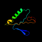
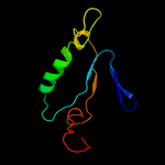
Top model
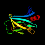
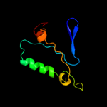
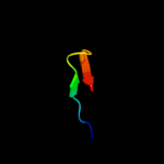
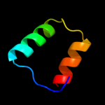
Image coloured by rainbow N → C terminus
Model (left) based on template c3dtdI_
Top template information
PDB header:structural genomics, unknown function
Chain: I: PDB Molecule:invasion-associated protein b;
PDBTitle: crystal structure of invasion associated protein b from bartonella2 henselae
Confidence and coverage
Confidence: 98.8% Coverage: 36%
126 residues ( 36% of your sequence) have been modelled with 98.8% confidence by the single highest scoring template.
You may wish to submit your sequence to Phyrealarm. This will automatically scan your sequence every week for new potential templates as they appear in the Phyre2 library.
3D viewing
Interactive 3D view in Jmol

Sequence analysis 

Secondary structure and disorder prediction 

   1........10.........20.........30.........40.........50.........60
Sequence  MRYRIFLLFFFALLPTSLVWAAPAQRAFSDWQVTCNNQNFCVARNTGDHNGLVMTLSRSA
Secondary structure 


SS confidence 


Disorder  ????


???????????


??


?
Disorder confidence 


   .........70.........80.........90.........100.........110.........120
Sequence  GAHTDAVLRIERGGLKSPEASEGEIAPRLLLDGEPLALSGDKWRISPWLLVTDDTATITA
Secondary structure 


SS confidence 


Disorder 

?


???????????


???????????


?


Disorder confidence 


   .........130.........140.........150.........160.........170.........180
Sequence  FLQMIQEGKAITLRDGDQTISLSGLKAALLFIDAQQKRVGSETAWIKKGDEPPLSVPPAP
Secondary structure 


SS confidence 


Disorder 


???


?
???


?


??
??
Disorder confidence 


   .........190.........200.........210.........220.........230.........240
Sequence  ALKEVAVVNPTPTPLSLEERNDLLDYGNWRMNGLRCSLDPLRREVNVTALTDDKALMMIS
Secondary structure 


SS confidence 


Disorder  ???
????
???????????


??
?
?????


Disorder confidence 


   .........250.........260.........270.........280.........290.........300
Sequence  CEAGAYNTIDLAWIVSRKKPLASRPVRLRLPFNNGQETNELELMNATFDEKSRELVTLAK
Secondary structure 


SS confidence 


Disorder 


??
?
?


???????????
??


Disorder confidence 


   .........310.........320.........330.........340.......
Sequence  GRGLSDCGIQARWRFDGQRFRLVRYAAEPTCDNWHGPDAWPTLWITR
Secondary structure 


SS confidence 


Disorder 


????????????


????
Disorder confidence 


Confidence Key
High(9)                    Low (0)
?Disordered
Alpha helix
Beta strand

Domain analysis 

Hover over an aligned region to see model and summary info

Please note, only up to the top 20 hits are modelled to reduce computer load

RankAligned region

PDB 3dtd chain I

3D model

Region: 24 - 156
Aligned: 126
Modelled: 128
Confidence: 98.8%
Identity: 20%
PDB header:structural genomics, unknown function
Chain: I: PDB Molecule:invasion-associated protein b;
PDBTitle: crystal structure of invasion associated protein b from bartonella2 henselae

Phyre2

PDB 1jke chain A

3D model

Region: 87 - 172
Aligned: 81
Modelled: 86
Confidence: 30.4%
Identity: 12%
Fold: DTD-like
Superfamily: DTD-like
Family: DTD-like

Phyre2

PDB 2byc chain A domain 1

3D model

Region: 25 - 37
Aligned: 13
Modelled: 13
Confidence: 18.5%
Identity: 31%
Fold: Ferredoxin-like
Superfamily: Acylphosphatase/BLUF domain-like
Family: BLUF domain

Phyre2

PDB 3ko7 chain E

3D model

Region: 87 - 172
Aligned: 81
Modelled: 86
Confidence: 18.5%
Identity: 12%
PDB header:hydrolase
Chain: E: PDB Molecule:d-tyrosyl-trna(tyr) deacylase;
PDBTitle: dtd from plasmodium falciparum in complex with d-lysine

Phyre2

PDB 1j7g chain A

3D model

Region: 87 - 172
Aligned: 81
Modelled: 86
Confidence: 17.4%
Identity: 12%
Fold: DTD-like
Superfamily: DTD-like
Family: DTD-like

Phyre2

PDB 2kb2 chain A

3D model

Region: 25 - 39
Aligned: 15
Modelled: 15
Confidence: 16.3%
Identity: 13%
PDB header:signaling protein, hydrolase regulator
Chain: A: PDB Molecule:blrp1;
PDBTitle: blrp1 bluf

Phyre2

PDB 2elc chain A domain 1

3D model

Region: 91 - 126
Aligned: 36
Modelled: 36
Confidence: 16.2%
Identity: 11%
Fold: Methionine synthase domain-like
Superfamily: Nucleoside phosphorylase/phosphoribosyltransferase N-terminal domain
Family: Nucleoside phosphorylase/phosphoribosyltransferase N-terminal domain

Phyre2

PDB 2hfn chain J

3D model

Region: 25 - 35
Aligned: 11
Modelled: 11
Confidence: 15.8%
Identity: 27%
PDB header:electron transport
Chain: J: PDB Molecule:synechocystis photoreceptor (slr1694);
PDBTitle: crystal structures of the synechocystis photoreceptor slr1694 reveal2 distinct structural states related to signaling

Phyre2

PDB 2okv chain C

3D model

Region: 87 - 172
Aligned: 80
Modelled: 86
Confidence: 14.2%
Identity: 14%
PDB header:hydrolase
Chain: C: PDB Molecule:probable d-tyrosyl-trna(tyr) deacylase 1;
PDBTitle: c-myc dna unwinding element binding protein

Phyre2

PDB 2o8r chain A domain 3

3D model

Region: 114 - 133
Aligned: 20
Modelled: 20
Confidence: 14.0%
Identity: 25%
Fold: Phospholipase D/nuclease
Superfamily: Phospholipase D/nuclease
Family: Polyphosphate kinase C-terminal domain

Phyre2

PDB 1x0p chain A domain 1

3D model

Region: 25 - 35
Aligned: 11
Modelled: 11
Confidence: 13.2%
Identity: 27%
Fold: Ferredoxin-like
Superfamily: Acylphosphatase/BLUF domain-like
Family: BLUF domain

Phyre2

PDB 1yrx chain A domain 1

3D model

Region: 25 - 35
Aligned: 11
Modelled: 11
Confidence: 12.0%
Identity: 27%
Fold: Ferredoxin-like
Superfamily: Acylphosphatase/BLUF domain-like
Family: BLUF domain

Phyre2

PDB 2wjv chain E

3D model

Region: 150 - 171
Aligned: 22
Modelled: 22
Confidence: 11.3%
Identity: 18%
PDB header:hydrolase
Chain: E: PDB Molecule:regulator of nonsense transcripts 2;
PDBTitle: crystal structure of the complex between human nonsense2 mediated decay factors upf1 and upf2

Phyre2

PDB 2dbo chain A

3D model

Region: 87 - 172
Aligned: 81
Modelled: 86
Confidence: 11.1%
Identity: 17%
PDB header:hydrolase
Chain: A: PDB Molecule:d-tyrosyl-trna(tyr) deacylase;
PDBTitle: crystal structure of d-tyr-trna(tyr) deacylase from aquifex aeolicus

Phyre2

PDB 2d32 chain A domain 1

3D model

Region: 1 - 16
Aligned: 16
Modelled: 16
Confidence: 10.0%
Identity: 19%
Fold: Glutamine synthetase/guanido kinase
Superfamily: Glutamine synthetase/guanido kinase
Family: Glutamate-cysteine ligase

Phyre2

PDB 3nzt chain A

3D model

Region: 1 - 17
Aligned: 17
Modelled: 17
Confidence: 9.9%
Identity: 18%
PDB header:ligase
Chain: A: PDB Molecule:glutamate--cysteine ligase;
PDBTitle: 2.0 angstrom crystal structure of glutamate--cysteine ligase (gsha)2 ftom francisella tularensis in complex with amp

Phyre2

PDB 1uou chain A domain 1

3D model

Region: 91 - 126
Aligned: 36
Modelled: 36
Confidence: 9.8%
Identity: 22%
Fold: Methionine synthase domain-like
Superfamily: Nucleoside phosphorylase/phosphoribosyltransferase N-terminal domain
Family: Nucleoside phosphorylase/phosphoribosyltransferase N-terminal domain

Phyre2

PDB 1brw chain A domain 1

3D model

Region: 91 - 126
Aligned: 36
Modelled: 36
Confidence: 9.1%
Identity: 17%
Fold: Methionine synthase domain-like
Superfamily: Nucleoside phosphorylase/phosphoribosyltransferase N-terminal domain
Family: Nucleoside phosphorylase/phosphoribosyltransferase N-terminal domain

Phyre2

PDB 2ov2 chain O

3D model

Region: 283 - 296
Aligned: 14
Modelled: 14
Confidence: 9.0%
Identity: 14%
PDB header:protein binding/transferase
Chain: O: PDB Molecule:serine/threonine-protein kinase pak 4;
PDBTitle: the crystal structure of the human rac3 in complex with the crib2 domain of human p21-activated kinase 4 (pak4)

Phyre2

PDB 1khd chain A domain 1

3D model

Region: 91 - 126
Aligned: 36
Modelled: 36
Confidence: 8.8%
Identity: 6%
Fold: Methionine synthase domain-like
Superfamily: Nucleoside phosphorylase/phosphoribosyltransferase N-terminal domain
Family: Nucleoside phosphorylase/phosphoribosyltransferase N-terminal domain

Phyre2
1

c3dtdI_
2

d1jkea_
3

d2byca1
4

c3ko7E_
5

d1j7ga_
6

c2kb2A_
7

d2elca1
8

c2hfnJ_
9

c2okvC_
10

d2o8ra3
11

d1x0pa1
12

d1yrxa1
13

c2wjvE_
14

c2dboA_
15

d2d32a1
16

c3nztA_
17

d1uoua1
18

d1brwa1
19

c2ov2O_
20

d1khda1
21


22


23


24


25


26


27


28


29


30


31


32


33


34


35


36


Detailed template information 

#
Template Alignment Coverage3D Model Confidence
% i.d. Template Information
1c3dtdI_


98.8 20 PDB header:structural genomics, unknown function
Chain: I: PDB Molecule:invasion-associated protein b;
PDBTitle: crystal structure of invasion associated protein b from bartonella2 henselae
2d1jkea_


30.4 12 Fold:DTD-like
Superfamily:DTD-like
Family:DTD-like
3d2byca1


18.5 31 Fold:Ferredoxin-like
Superfamily:Acylphosphatase/BLUF domain-like
Family:BLUF domain
4c3ko7E_


18.5 12 PDB header:hydrolase
Chain: E: PDB Molecule:d-tyrosyl-trna(tyr) deacylase;
PDBTitle: dtd from plasmodium falciparum in complex with d-lysine
5d1j7ga_


17.4 12 Fold:DTD-like
Superfamily:DTD-like
Family:DTD-like
6c2kb2A_


16.3 13 PDB header:signaling protein, hydrolase regulator
Chain: A: PDB Molecule:blrp1;
PDBTitle: blrp1 bluf
7d2elca1


16.2 11 Fold:Methionine synthase domain-like
Superfamily:Nucleoside phosphorylase/phosphoribosyltransferase N-terminal domain
Family:Nucleoside phosphorylase/phosphoribosyltransferase N-terminal domain
8c2hfnJ_


15.8 27 PDB header:electron transport
Chain: J: PDB Molecule:synechocystis photoreceptor (slr1694);
PDBTitle: crystal structures of the synechocystis photoreceptor slr1694 reveal2 distinct structural states related to signaling
9c2okvC_


14.2 14 PDB header:hydrolase
Chain: C: PDB Molecule:probable d-tyrosyl-trna(tyr) deacylase 1;
PDBTitle: c-myc dna unwinding element binding protein
10d2o8ra3


14.0 25 Fold:Phospholipase D/nuclease
Superfamily:Phospholipase D/nuclease
Family:Polyphosphate kinase C-terminal domain
11d1x0pa1


13.2 27 Fold:Ferredoxin-like
Superfamily:Acylphosphatase/BLUF domain-like
Family:BLUF domain
12d1yrxa1


12.0 27 Fold:Ferredoxin-like
Superfamily:Acylphosphatase/BLUF domain-like
Family:BLUF domain
13c2wjvE_


11.3 18 PDB header:hydrolase
Chain: E: PDB Molecule:regulator of nonsense transcripts 2;
PDBTitle: crystal structure of the complex between human nonsense2 mediated decay factors upf1 and upf2
14c2dboA_


11.1 17 PDB header:hydrolase
Chain: A: PDB Molecule:d-tyrosyl-trna(tyr) deacylase;
PDBTitle: crystal structure of d-tyr-trna(tyr) deacylase from aquifex aeolicus
15d2d32a1


10.0 19 Fold:Glutamine synthetase/guanido kinase
Superfamily:Glutamine synthetase/guanido kinase
Family:Glutamate-cysteine ligase
16c3nztA_


9.9 18 PDB header:ligase
Chain: A: PDB Molecule:glutamate--cysteine ligase;
PDBTitle: 2.0 angstrom crystal structure of glutamate--cysteine ligase (gsha)2 ftom francisella tularensis in complex with amp
17d1uoua1


9.8 22 Fold:Methionine synthase domain-like
Superfamily:Nucleoside phosphorylase/phosphoribosyltransferase N-terminal domain
Family:Nucleoside phosphorylase/phosphoribosyltransferase N-terminal domain
18d1brwa1


9.1 17 Fold:Methionine synthase domain-like
Superfamily:Nucleoside phosphorylase/phosphoribosyltransferase N-terminal domain
Family:Nucleoside phosphorylase/phosphoribosyltransferase N-terminal domain
19c2ov2O_


9.0 14 PDB header:protein binding/transferase
Chain: O: PDB Molecule:serine/threonine-protein kinase pak 4;
PDBTitle: the crystal structure of the human rac3 in complex with the crib2 domain of human p21-activated kinase 4 (pak4)
20d1khda1


8.8 6 Fold:Methionine synthase domain-like
Superfamily:Nucleoside phosphorylase/phosphoribosyltransferase N-terminal domain
Family:Nucleoside phosphorylase/phosphoribosyltransferase N-terminal domain
21d1v8ga1


not modelled 8.7 11 Fold:Methionine synthase domain-like
Superfamily:Nucleoside phosphorylase/phosphoribosyltransferase N-terminal domain
Family:Nucleoside phosphorylase/phosphoribosyltransferase N-terminal domain
22d1u2ca2


not modelled 7.2 29 Fold:Dystroglycan, domain 2
Superfamily:Dystroglycan, domain 2
Family:Dystroglycan, domain 2
23d2tpta1


not modelled 7.2 23 Fold:Methionine synthase domain-like
Superfamily:Nucleoside phosphorylase/phosphoribosyltransferase N-terminal domain
Family:Nucleoside phosphorylase/phosphoribosyltransferase N-terminal domain
24c2odbB_


not modelled 7.2 23 PDB header:protein binding
Chain: B: PDB Molecule:serine/threonine-protein kinase pak 6;
PDBTitle: the crystal structure of human cdc42 in complex with the crib domain2 of human p21-activated kinase 6 (pak6)
25d1j26a_


not modelled 6.8 22 Fold:dsRBD-like
Superfamily:Peptidyl-tRNA hydrolase domain-like
Family:Peptidyl-tRNA hydrolase domain
26c2o8rA_


not modelled 6.7 24 PDB header:transferase
Chain: A: PDB Molecule:polyphosphate kinase;
PDBTitle: crystal structure of polyphosphate kinase from2 porphyromonas gingivalis
27c2bs5A_


not modelled 6.6 28 PDB header:sugar binding protein
Chain: A: PDB Molecule:fucose-binding lectin protein;
PDBTitle: lectin from ralstonia solanacearum complexed with 2-2 fucosyllactose
28d1o17a1


not modelled 6.4 8 Fold:Methionine synthase domain-like
Superfamily:Nucleoside phosphorylase/phosphoribosyltransferase N-terminal domain
Family:Nucleoside phosphorylase/phosphoribosyltransferase N-terminal domain
29d1f9fa_


not modelled 6.4 15 Fold:Ferredoxin-like
Superfamily:Viral DNA-binding domain
Family:Viral DNA-binding domain
30c3ln6A_


not modelled 6.1 31 PDB header:ligase
Chain: A: PDB Molecule:glutathione biosynthesis bifunctional protein gshab;
PDBTitle: crystal structure of a bifunctional glutathione synthetase from2 streptococcus agalactiae
31d1ut9a1


not modelled 6.1 6 Fold:alpha/alpha toroid
Superfamily:Six-hairpin glycosidases
Family:Cellulases catalytic domain
32c3ln7A_


not modelled 6.1 44 PDB header:ligase
Chain: A: PDB Molecule:glutathione biosynthesis bifunctional protein gshab;
PDBTitle: crystal structure of a bifunctional glutathione synthetase from2 pasteurella multocida
33c1wz5A_


not modelled 6.0 78 PDB header:toxin
Chain: A: PDB Molecule:potassium channel blocking toxin 1;
PDBTitle: solution structure of pi1-3p
34c1f3mB_


not modelled 6.0 21 PDB header:transferase
Chain: B: PDB Molecule:serine/threonine-protein kinase pak-alpha;
PDBTitle: crystal structure of human serine/threonine kinase pak1
35c1w8xN_


not modelled 5.7 14 PDB header:virus
Chain: N: PDB Molecule:protein p31;
PDBTitle: structural analysis of prd1
36c1e0aB_


not modelled 5.6 14 PDB header:signalling protein
Chain: B: PDB Molecule:serine/threonine-protein kinase pak-alpha;
PDBTitle: cdc42 complexed with the gtpase binding domain of p212 activated kinase

Binding site prediction 

Due to computational demand, binding site predictions are not run for batch jobs

If you want to predict binding sites, please manually submit your model of choice to 3DLigandSite

Transmembrane helix prediction 

Transmembrane helices have been predicted in your sequence to adopt the topology shown below


Phyre is for academic use only

Please cite: Protein structure prediction on the web: a case study using the Phyre server
Kelley LA and Sternberg MJE. Nature Protocols 4, 363 - 371 (2009) [pdf] [Import into BibTeX]
 
If you use the binding site predictions from 3DLigandSite, please also cite:
3DLigandSite: predicting ligand-binding sites using similar structures.
Wass MN, Kelley LA and Sternberg MJ Nucleic Acids Research 38, W469-73 (2010) [PubMed]
 
© Structural Bioinformatics Group
Imperial College London
Lawrence Kelley, Benjamin Jefferys
Disclaimer
Terms and Conditions
Component software
Template detection: HHpred 1.51
Secondary structure prediction: Psi-pred 2.5
Disorder prediction: Disopred 2.4
Transmembrane prediction: Memsat_SVM
Multi-template modelling and ab initio: Poing 1.0