| 1 |
|
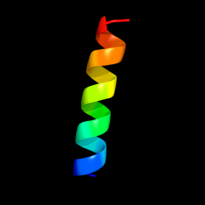
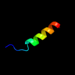
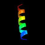
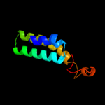
PDB 1zxa chain B
Region: 5 - 22
Aligned: 18
Modelled: 18
Confidence: 30.3%
Identity: 56%
PDB header:transferase
Chain: B: PDB Molecule:cgmp-dependent protein kinase 1, alpha isozyme;
PDBTitle: solution structure of the coiled-coil domain of cgmp-2 dependent protein kinase ia
Phyre2
| 2 |
|
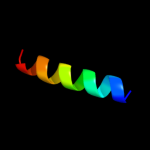
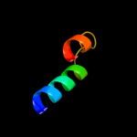
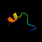
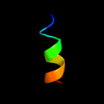
PDB 1t3j chain A
Region: 16 - 51
Aligned: 36
Modelled: 36
Confidence: 20.6%
Identity: 31%
PDB header:membrane protein
Chain: A: PDB Molecule:mitofusin 1;
PDBTitle: mitofusin domain hr2 v686m/i708m mutant
Phyre2
| 3 |
|
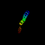
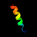
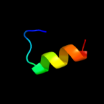
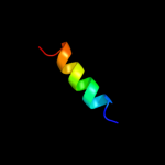
PDB 1r2a chain A
Region: 64 - 83
Aligned: 20
Modelled: 20
Confidence: 18.9%
Identity: 40%
Fold: Dimerization-anchoring domain of cAMP-dependent PK regulatory subunit
Superfamily: Dimerization-anchoring domain of cAMP-dependent PK regulatory subunit
Family: Dimerization-anchoring domain of cAMP-dependent PK regulatory subunit
Phyre2
| 4 |
|
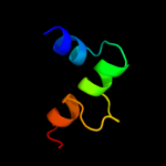
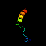
PDB 2b8i chain A
Region: 1 - 25
Aligned: 25
Modelled: 25
Confidence: 17.7%
Identity: 40%
PDB header:lipid binding protein
Chain: A: PDB Molecule:pas factor;
PDBTitle: crystal structure and functional studies reveal that pas2 factor from vibrio vulnificus is a novel member of the3 saposin-fold family
Phyre2
| 5 |
|
PDB 2hwn chain A domain 1
Region: 65 - 83
Aligned: 19
Modelled: 19
Confidence: 16.3%
Identity: 42%
Fold: Dimerization-anchoring domain of cAMP-dependent PK regulatory subunit
Superfamily: Dimerization-anchoring domain of cAMP-dependent PK regulatory subunit
Family: Dimerization-anchoring domain of cAMP-dependent PK regulatory subunit
Phyre2
| 6 |
|
PDB 2qsh chain A
Region: 7 - 43
Aligned: 31
Modelled: 37
Confidence: 12.2%
Identity: 23%
PDB header:dna binding protein/dna
Chain: A: PDB Molecule:dna repair protein rad4;
PDBTitle: crystal structure of rad4-rad23 bound to a mismatch dna
Phyre2
| 7 |
|
PDB 1p9i chain A
Region: 12 - 29
Aligned: 18
Modelled: 18
Confidence: 9.2%
Identity: 56%
PDB header:unknown function
Chain: A: PDB Molecule:cortexillin i/gcn4 hybrid peptide;
PDBTitle: coiled-coil x-ray structure at 1.17 a resolution
Phyre2
| 8 |
|
PDB 1v2z chain A
Region: 37 - 52
Aligned: 16
Modelled: 16
Confidence: 8.5%
Identity: 31%
Fold: KaiA/RbsU domain
Superfamily: KaiA/RbsU domain
Family: Circadian clock protein KaiA, C-terminal domain
Phyre2
| 9 |
|
PDB 1r8j chain A domain 1
Region: 37 - 52
Aligned: 16
Modelled: 16
Confidence: 7.3%
Identity: 31%
Fold: KaiA/RbsU domain
Superfamily: KaiA/RbsU domain
Family: Circadian clock protein KaiA, C-terminal domain
Phyre2
| 10 |
|
PDB 1o0l chain A
Region: 54 - 84
Aligned: 30
Modelled: 31
Confidence: 6.5%
Identity: 37%
Fold: Toxins' membrane translocation domains
Superfamily: Bcl-2 inhibitors of programmed cell death
Family: Bcl-2 inhibitors of programmed cell death
Phyre2
| 11 |
|
PDB 3cqc chain B
Region: 6 - 84
Aligned: 74
Modelled: 79
Confidence: 6.2%
Identity: 19%
PDB header:protein transport
Chain: B: PDB Molecule:nuclear pore complex protein nup133;
PDBTitle: nucleoporin nup107/nup133 interaction complex
Phyre2
| 12 |
|
PDB 1peh chain A
Region: 16 - 27
Aligned: 12
Modelled: 12
Confidence: 6.1%
Identity: 58%
PDB header:nucleotidyltransferase
Chain: A: PDB Molecule:pepnh1;
PDBTitle: nmr structure of the membrane-binding domain of ctp2 phosphocholine cytidylyltransferase, 10 structures
Phyre2
| 13 |
|
PDB 1y14 chain B domain 2
Region: 38 - 54
Aligned: 17
Modelled: 17
Confidence: 5.5%
Identity: 35%
Fold: Dodecin subunit-like
Superfamily: N-terminal, heterodimerisation domain of RBP7 (RpoE)
Family: N-terminal, heterodimerisation domain of RBP7 (RpoE)
Phyre2