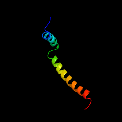
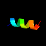
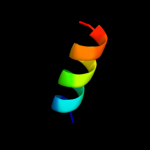
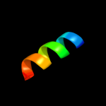
Phyre Home Retrieve Phyre Job Id
Emaill.a.kelley@imperial.ac.uk
DescriptionP0A843
DateWed Jan 25 15:20:18 GMT 2012
Unique Job ID34d777637fc236cf

Summary 

Top model
Image coloured by rainbow N → C terminus
Model (left) based on template c2l16A_
Top template information
PDB header:protein transport
Chain: A: PDB Molecule:sec-independent protein translocase protein tatad;
PDBTitle: solution structure of bacillus subtilits tatad protein in dpc micelles
Confidence and coverage
Confidence: 99.8% Coverage: 76%
51 residues ( 76% of your sequence) have been modelled with 99.8% confidence by the single highest scoring template.
3D viewing
Interactive 3D view in Jmol

Sequence analysis 

Secondary structure and disorder prediction 

   1........10.........20.........30.........40.........50.........60
Sequence  MGEISITKLLVVAALVVLLFGTKKLRTLGGDLGAAIKGFKKAMNDDDAAAKKGADVDLQA
Secondary structure 


SS confidence 


Disorder  ?????


??????????????????
Disorder confidence 


   .......
Sequence  EKLSHKE
Secondary structure 


SS confidence 


Disorder  ???????
Disorder confidence 


Confidence Key
High(9)                    Low (0)
?Disordered
Alpha helix
Beta strand

Domain analysis 

Hover over an aligned region to see model and summary info

Please note, only up to the top 20 hits are modelled to reduce computer load

RankAligned region

PDB 2l16 chain A

3D model

Region: 1 - 51
Aligned: 51
Modelled: 51
Confidence: 99.8%
Identity: 25%
PDB header:protein transport
Chain: A: PDB Molecule:sec-independent protein translocase protein tatad;
PDBTitle: solution structure of bacillus subtilits tatad protein in dpc micelles

Phyre2

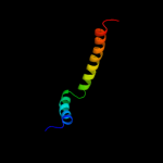
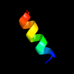
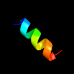
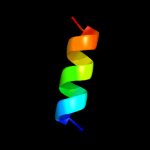
PDB 2cqq chain A domain 1

3D model

Region: 12 - 52
Aligned: 41
Modelled: 41
Confidence: 18.5%
Identity: 12%
Fold: DNA/RNA-binding 3-helical bundle
Superfamily: Homeodomain-like
Family: Myb/SANT domain

Phyre2

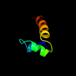
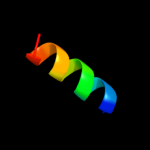
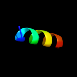
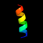
PDB 2axt chain I domain 1

3D model

Region: 9 - 21
Aligned: 13
Modelled: 13
Confidence: 15.0%
Identity: 23%
Fold: Single transmembrane helix
Superfamily: Photosystem II reaction center protein I, PsbI
Family: PsbI-like

Phyre2

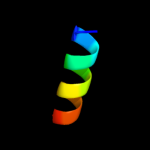
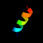
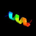
PDB 2axt chain L domain 1

3D model

Region: 6 - 19
Aligned: 14
Modelled: 14
Confidence: 9.2%
Identity: 29%
Fold: Single transmembrane helix
Superfamily: Photosystem II reaction center protein L, PsbL
Family: PsbL-like

Phyre2

PDB 3a0h chain L

3D model

Region: 6 - 19
Aligned: 14
Modelled: 14
Confidence: 9.2%
Identity: 29%
PDB header:electron transport
Chain: L: PDB Molecule:photosystem ii reaction center protein l;
PDBTitle: crystal structure of i-substituted photosystem ii complex

Phyre2

PDB 3bz1 chain L

3D model

Region: 6 - 19
Aligned: 14
Modelled: 14
Confidence: 9.2%
Identity: 29%
PDB header:electron transport
Chain: L: PDB Molecule:photosystem ii reaction center protein l;
PDBTitle: crystal structure of cyanobacterial photosystem ii (part 12 of 2). this file contains first monomer of psii dimer

Phyre2

PDB 3prq chain L

3D model

Region: 6 - 19
Aligned: 14
Modelled: 14
Confidence: 9.2%
Identity: 29%
PDB header:photosynthesis
Chain: L: PDB Molecule:photosystem ii reaction center protein l;
PDBTitle: crystal structure of cyanobacterial photosystem ii in complex with2 terbutryn (part 1 of 2). this file contains first monomer of psii3 dimer

Phyre2

PDB 2axt chain L

3D model

Region: 6 - 19
Aligned: 14
Modelled: 14
Confidence: 9.2%
Identity: 29%
PDB header:electron transport
Chain: L: PDB Molecule:photosystem ii reaction center l protein;
PDBTitle: crystal structure of photosystem ii from thermosynechococcus elongatus

Phyre2

PDB 3prr chain L

3D model

Region: 6 - 19
Aligned: 14
Modelled: 14
Confidence: 9.2%
Identity: 29%
PDB header:photosynthesis
Chain: L: PDB Molecule:photosystem ii reaction center protein l;
PDBTitle: crystal structure of cyanobacterial photosystem ii in complex with2 terbutryn (part 2 of 2). this file contains second monomer of psii3 dimer

Phyre2

PDB 3arc chain L

3D model

Region: 6 - 19
Aligned: 14
Modelled: 14
Confidence: 9.2%
Identity: 29%
PDB header:electron transport, photosynthesis
Chain: L: PDB Molecule:photosystem ii reaction center protein l;
PDBTitle: crystal structure of oxygen-evolving photosystem ii at 1.9 angstrom2 resolution

Phyre2

PDB 3bz2 chain L

3D model

Region: 6 - 19
Aligned: 14
Modelled: 14
Confidence: 9.2%
Identity: 29%
PDB header:electron transport
Chain: L: PDB Molecule:photosystem ii reaction center protein l;
PDBTitle: crystal structure of cyanobacterial photosystem ii (part 22 of 2). this file contains second monomer of psii dimer

Phyre2

PDB 3a0b chain L

3D model

Region: 6 - 19
Aligned: 14
Modelled: 14
Confidence: 9.2%
Identity: 29%
PDB header:electron transport
Chain: L: PDB Molecule:photosystem ii reaction center protein l;
PDBTitle: crystal structure of br-substituted photosystem ii complex

Phyre2

PDB 3kzi chain L

3D model

Region: 6 - 19
Aligned: 14
Modelled: 14
Confidence: 9.2%
Identity: 29%
PDB header:electron transport
Chain: L: PDB Molecule:photosystem ii reaction center protein l;
PDBTitle: crystal structure of monomeric form of cyanobacterial photosystem ii

Phyre2

PDB 1s5l chain L

3D model

Region: 6 - 19
Aligned: 14
Modelled: 14
Confidence: 9.2%
Identity: 29%
PDB header:photosynthesis
Chain: L: PDB Molecule:photosystem ii reaction center l protein;
PDBTitle: architecture of the photosynthetic oxygen evolving center

Phyre2

PDB 3a0b chain L

3D model

Region: 6 - 19
Aligned: 14
Modelled: 14
Confidence: 9.2%
Identity: 29%
PDB header:electron transport
Chain: L: PDB Molecule:photosystem ii reaction center protein l;
PDBTitle: crystal structure of br-substituted photosystem ii complex

Phyre2

PDB 1s5l chain L

3D model

Region: 6 - 19
Aligned: 14
Modelled: 14
Confidence: 9.2%
Identity: 29%
PDB header:photosynthesis
Chain: L: PDB Molecule:photosystem ii reaction center l protein;
PDBTitle: architecture of the photosynthetic oxygen evolving center

Phyre2

PDB 2axt chain L

3D model

Region: 6 - 19
Aligned: 14
Modelled: 14
Confidence: 9.2%
Identity: 29%
PDB header:electron transport
Chain: L: PDB Molecule:photosystem ii reaction center l protein;
PDBTitle: crystal structure of photosystem ii from thermosynechococcus elongatus

Phyre2

PDB 3a0h chain L

3D model

Region: 6 - 19
Aligned: 14
Modelled: 14
Confidence: 9.2%
Identity: 29%
PDB header:electron transport
Chain: L: PDB Molecule:photosystem ii reaction center protein l;
PDBTitle: crystal structure of i-substituted photosystem ii complex

Phyre2

PDB 2axt chain J domain 1

3D model

Region: 9 - 22
Aligned: 14
Modelled: 14
Confidence: 8.3%
Identity: 14%
Fold: Single transmembrane helix
Superfamily: Photosystem II reaction center protein J, PsbJ
Family: PsbJ-like

Phyre2

PDB 3arc chain L

3D model

Region: 6 - 19
Aligned: 14
Modelled: 14
Confidence: 8.1%
Identity: 29%
PDB header:electron transport, photosynthesis
Chain: L: PDB Molecule:photosystem ii reaction center protein l;
PDBTitle: crystal structure of oxygen-evolving photosystem ii at 1.9 angstrom2 resolution

Phyre2
1

c2l16A_
2

d2cqqa1
3

d2axti1
4

d2axtl1
5

c3a0hl_
6

c3bz1L_
7

c3prqL_
8

c2axtl_
9

c3prrL_
10

c3arcL_
11

c3bz2L_
12

c3a0bl_
13

c3kziL_
14

c1s5ll_
15

c3a0bL_
16

c1s5lL_
17

c2axtL_
18

c3a0hL_
19

d2axtj1
20

c3arcl_
21


22


23


Detailed template information 

#
Template Alignment Coverage3D Model Confidence
% i.d. Template Information
1c2l16A_


99.8 25 PDB header:protein transport
Chain: A: PDB Molecule:sec-independent protein translocase protein tatad;
PDBTitle: solution structure of bacillus subtilits tatad protein in dpc micelles
2d2cqqa1


18.5 12 Fold:DNA/RNA-binding 3-helical bundle
Superfamily:Homeodomain-like
Family:Myb/SANT domain
3d2axti1


15.0 23 Fold:Single transmembrane helix
Superfamily:Photosystem II reaction center protein I, PsbI
Family:PsbI-like
4d2axtl1


9.2 29 Fold:Single transmembrane helix
Superfamily:Photosystem II reaction center protein L, PsbL
Family:PsbL-like
5c3a0hl_


9.2 29 PDB header:electron transport
Chain: L: PDB Molecule:photosystem ii reaction center protein l;
PDBTitle: crystal structure of i-substituted photosystem ii complex
6c3bz1L_


9.2 29 PDB header:electron transport
Chain: L: PDB Molecule:photosystem ii reaction center protein l;
PDBTitle: crystal structure of cyanobacterial photosystem ii (part 12 of 2). this file contains first monomer of psii dimer
7c3prqL_


9.2 29 PDB header:photosynthesis
Chain: L: PDB Molecule:photosystem ii reaction center protein l;
PDBTitle: crystal structure of cyanobacterial photosystem ii in complex with2 terbutryn (part 1 of 2). this file contains first monomer of psii3 dimer
8c2axtl_


9.2 29 PDB header:electron transport
Chain: L: PDB Molecule:photosystem ii reaction center l protein;
PDBTitle: crystal structure of photosystem ii from thermosynechococcus elongatus
9c3prrL_


9.2 29 PDB header:photosynthesis
Chain: L: PDB Molecule:photosystem ii reaction center protein l;
PDBTitle: crystal structure of cyanobacterial photosystem ii in complex with2 terbutryn (part 2 of 2). this file contains second monomer of psii3 dimer
10c3arcL_


9.2 29 PDB header:electron transport, photosynthesis
Chain: L: PDB Molecule:photosystem ii reaction center protein l;
PDBTitle: crystal structure of oxygen-evolving photosystem ii at 1.9 angstrom2 resolution
11c3bz2L_


9.2 29 PDB header:electron transport
Chain: L: PDB Molecule:photosystem ii reaction center protein l;
PDBTitle: crystal structure of cyanobacterial photosystem ii (part 22 of 2). this file contains second monomer of psii dimer
12c3a0bl_


9.2 29 PDB header:electron transport
Chain: L: PDB Molecule:photosystem ii reaction center protein l;
PDBTitle: crystal structure of br-substituted photosystem ii complex
13c3kziL_


9.2 29 PDB header:electron transport
Chain: L: PDB Molecule:photosystem ii reaction center protein l;
PDBTitle: crystal structure of monomeric form of cyanobacterial photosystem ii
14c1s5ll_


9.2 29 PDB header:photosynthesis
Chain: L: PDB Molecule:photosystem ii reaction center l protein;
PDBTitle: architecture of the photosynthetic oxygen evolving center
15c3a0bL_


9.2 29 PDB header:electron transport
Chain: L: PDB Molecule:photosystem ii reaction center protein l;
PDBTitle: crystal structure of br-substituted photosystem ii complex
16c1s5lL_


9.2 29 PDB header:photosynthesis
Chain: L: PDB Molecule:photosystem ii reaction center l protein;
PDBTitle: architecture of the photosynthetic oxygen evolving center
17c2axtL_


9.2 29 PDB header:electron transport
Chain: L: PDB Molecule:photosystem ii reaction center l protein;
PDBTitle: crystal structure of photosystem ii from thermosynechococcus elongatus
18c3a0hL_


9.2 29 PDB header:electron transport
Chain: L: PDB Molecule:photosystem ii reaction center protein l;
PDBTitle: crystal structure of i-substituted photosystem ii complex
19d2axtj1


8.3 14 Fold:Single transmembrane helix
Superfamily:Photosystem II reaction center protein J, PsbJ
Family:PsbJ-like
20c3arcl_


8.1 29 PDB header:electron transport, photosynthesis
Chain: L: PDB Molecule:photosystem ii reaction center protein l;
PDBTitle: crystal structure of oxygen-evolving photosystem ii at 1.9 angstrom2 resolution
21c1by0A_


not modelled 8.0 47 PDB header:rna binding protein
Chain: A: PDB Molecule:protein (hepatitis delta antigen);
PDBTitle: n-terminal leucine-repeat region of hepatitis delta antigen
22c1a92B_


not modelled 6.3 41 PDB header:leucine zipper
Chain: B: PDB Molecule:delta antigen;
PDBTitle: oligomerization domain of hepatitis delta antigen
23c2kdcC_


not modelled 5.9 9 PDB header:transferase
Chain: C: PDB Molecule:diacylglycerol kinase;
PDBTitle: nmr solution structure of e. coli diacylglycerol kinase2 (dagk) in dpc micelles

Binding site prediction 

Due to computational demand, binding site predictions are not run for batch jobs

If you want to predict binding sites, please manually submit your model of choice to 3DLigandSite


Phyre is for academic use only

Please cite: Protein structure prediction on the web: a case study using the Phyre server
Kelley LA and Sternberg MJE. Nature Protocols 4, 363 - 371 (2009) [pdf] [Import into BibTeX]
 
If you use the binding site predictions from 3DLigandSite, please also cite:
3DLigandSite: predicting ligand-binding sites using similar structures.
Wass MN, Kelley LA and Sternberg MJ Nucleic Acids Research 38, W469-73 (2010) [PubMed]
 
© Structural Bioinformatics Group
Imperial College London
Lawrence Kelley, Benjamin Jefferys
Disclaimer
Terms and Conditions
Component software
Template detection: HHpred 1.51
Secondary structure prediction: Psi-pred 2.5
Disorder prediction: Disopred 2.4
Transmembrane prediction: Memsat_SVM
Multi-template modelling and ab initio: Poing 1.0