Phyre Home Retrieve Phyre Job Id
Emaill.a.kelley@imperial.ac.uk
DescriptionP37194
DateThu Jan 5 11:55:00 GMT 2012
Unique Job ID33ca6ece8a835ac5

Summary 

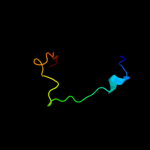
Top model
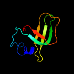
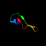
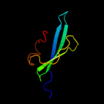
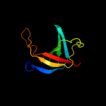
Image coloured by rainbow N → C terminus
Model (left) based on template c1j6qA_
Top template information
PDB header:chaperone
Chain: A: PDB Molecule:cytochrome c maturation protein e;
PDBTitle: solution structure and characterization of the heme2 chaperone ccme
Confidence and coverage
Confidence: 82.3% Coverage: 41%
77 residues ( 41% of your sequence) have been modelled with 82.3% confidence by the single highest scoring template.
You may wish to submit your sequence to Phyrealarm. This will automatically scan your sequence every week for new potential templates as they appear in the Phyre2 library.
Please note: You must be registered and logged in to use Phyrealarm.
3D viewing
Interactive 3D view in Jmol

Sequence analysis 

Secondary structure and disorder prediction 

   1........10.........20.........30.........40.........50.........60
Sequence  MNMTKGALILSLSFLLAACSSIPQNIKGNNQPDIQKSFVAVHNQPGLYVGQQARFGGKVI
Secondary structure 


SS confidence 


Disorder  ???


??????????????????


???

??


Disorder confidence 


   .........70.........80.........90.........100.........110.........120
Sequence  NVINGKTDTLLEISVLPLDSYAKPDIEANYQGRLLARQSGFLDPVNYRNHFVTILGTIQG
Secondary structure 


SS confidence 


Disorder 


??????????


???
Disorder confidence 


   .........130.........140.........150.........160.........170.........180
Sequence  EQPGFINKVPYNFLEVNMQGIQVWHLREVVNTTYNLWDYGYGAFWPEPGWGAPYYTNAVS
Secondary structure 


SS confidence 


Disorder  ??


??


?
?????


Disorder confidence 


   ........
Sequence  QVTPELVK
Secondary structure 


SS confidence 


Disorder 
???????
Disorder confidence 


Confidence Key
High(9)                    Low (0)
?Disordered
Alpha helix
Beta strand

Domain analysis 

Hover over an aligned region to see model and summary info

Please note, only up to the top 20 hits are modelled to reduce computer load

RankAligned region

PDB 1j6q chain A

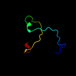
3D model

Region: 32 - 120
Aligned: 77
Modelled: 78
Confidence: 82.3%
Identity: 17%
PDB header:chaperone
Chain: A: PDB Molecule:cytochrome c maturation protein e;
PDBTitle: solution structure and characterization of the heme2 chaperone ccme

Phyre2

PDB 1j6q chain A

3D model

Region: 32 - 120
Aligned: 77
Modelled: 78
Confidence: 82.3%
Identity: 17%
Fold: OB-fold
Superfamily: Heme chaperone CcmE
Family: Heme chaperone CcmE

Phyre2

PDB 1sr3 chain A

3D model

Region: 32 - 119
Aligned: 75
Modelled: 88
Confidence: 66.0%
Identity: 21%
Fold: OB-fold
Superfamily: Heme chaperone CcmE
Family: Heme chaperone CcmE

Phyre2

PDB 2kct chain A

3D model

Region: 42 - 119
Aligned: 67
Modelled: 78
Confidence: 49.5%
Identity: 16%
PDB header:chaperone
Chain: A: PDB Molecule:cytochrome c-type biogenesis protein ccme;
PDBTitle: solution nmr structure of the ob-fold domain of heme2 chaperone ccme from desulfovibrio vulgaris. northeast3 structural genomics target dvr115g.

Phyre2

PDB 2r9q chain D

3D model

Region: 69 - 104
Aligned: 36
Modelled: 36
Confidence: 40.9%
Identity: 22%
PDB header:hydrolase
Chain: D: PDB Molecule:2'-deoxycytidine 5'-triphosphate deaminase;
PDBTitle: crystal structure of 2'-deoxycytidine 5'-triphosphate deaminase from2 agrobacterium tumefaciens

Phyre2

PDB 3h43 chain F

3D model

Region: 89 - 114
Aligned: 26
Modelled: 26
Confidence: 27.8%
Identity: 19%
PDB header:hydrolase
Chain: F: PDB Molecule:proteasome-activating nucleotidase;
PDBTitle: n-terminal domain of the proteasome-activating nucleotidase2 of methanocaldococcus jannaschii

Phyre2

PDB 2wkd chain A

3D model

Region: 96 - 137
Aligned: 32
Modelled: 42
Confidence: 27.7%
Identity: 25%
PDB header:dna binding protein
Chain: A: PDB Molecule:orf34p2;
PDBTitle: crystal structure of a double ile-to-met mutant of protein2 orf34 from lactococcus phage p2

Phyre2

PDB 1n9w chain A domain 1

3D model

Region: 43 - 73
Aligned: 31
Modelled: 31
Confidence: 26.4%
Identity: 16%
Fold: OB-fold
Superfamily: Nucleic acid-binding proteins
Family: Anticodon-binding domain

Phyre2

PDB 2jwy chain A

3D model

Region: 42 - 127
Aligned: 86
Modelled: 86
Confidence: 25.2%
Identity: 13%
PDB header:lipoprotein
Chain: A: PDB Molecule:uncharacterized lipoprotein yaji;
PDBTitle: solution nmr structure of uncharacterized lipoprotein yaji from2 escherichia coli. northeast structural genomics target er540

Phyre2

PDB 2wg6 chain L

3D model

Region: 89 - 114
Aligned: 26
Modelled: 26
Confidence: 24.9%
Identity: 8%
PDB header:transcription,hydrolase
Chain: L: PDB Molecule:general control protein gcn4,
PDBTitle: proteasome-activating nucleotidase (pan) n-domain (57-134)2 from archaeoglobus fulgidus fused to gcn4, p61a mutant

Phyre2

PDB 2z14 chain A

3D model

Region: 47 - 118
Aligned: 72
Modelled: 72
Confidence: 24.6%
Identity: 17%
PDB header:signaling protein
Chain: A: PDB Molecule:ef-hand domain-containing family member c2;
PDBTitle: crystal structure of the n-terminal duf1126 in human ef-2 hand domain containing 2 protein

Phyre2

PDB 2wp8 chain J

3D model

Region: 57 - 122
Aligned: 65
Modelled: 66
Confidence: 23.5%
Identity: 28%
PDB header:hydrolase
Chain: J: PDB Molecule:exosome complex exonuclease dis3;
PDBTitle: yeast rrp44 nuclease

Phyre2

PDB 2pi2 chain E domain 1

3D model

Region: 42 - 120
Aligned: 61
Modelled: 79
Confidence: 20.2%
Identity: 15%
Fold: OB-fold
Superfamily: Nucleic acid-binding proteins
Family: Single strand DNA-binding domain, SSB

Phyre2

PDB 1l0w chain A domain 1

3D model

Region: 46 - 73
Aligned: 28
Modelled: 28
Confidence: 18.1%
Identity: 18%
Fold: OB-fold
Superfamily: Nucleic acid-binding proteins
Family: Anticodon-binding domain

Phyre2

PDB 1wyd chain B

3D model

Region: 41 - 143
Aligned: 83
Modelled: 89
Confidence: 17.9%
Identity: 12%
PDB header:ligase
Chain: B: PDB Molecule:hypothetical aspartyl-trna synthetase;
PDBTitle: crystal structure of aspartyl-trna synthetase from sulfolobus tokodaii

Phyre2

PDB 2vnu chain D domain 1

3D model

Region: 93 - 122
Aligned: 30
Modelled: 30
Confidence: 17.9%
Identity: 33%
Fold: OB-fold
Superfamily: Nucleic acid-binding proteins
Family: Cold shock DNA-binding domain-like

Phyre2

PDB 1t3t chain A domain 6

3D model

Region: 88 - 126
Aligned: 39
Modelled: 39
Confidence: 16.8%
Identity: 18%
Fold: PurM C-terminal domain-like
Superfamily: PurM C-terminal domain-like
Family: PurM C-terminal domain-like

Phyre2

PDB 1vqw chain B

3D model

Region: 16 - 116
Aligned: 99
Modelled: 100
Confidence: 14.4%
Identity: 14%
PDB header:structural genomics, unknown function
Chain: B: PDB Molecule:protein with similarity to flavin-containing
PDBTitle: crystal structure of a protein with similarity to flavin-2 containing monooxygenases and to mammalian dimethylalanine3 monooxygenases

Phyre2

PDB 1yrl chain D

3D model

Region: 81 - 116
Aligned: 35
Modelled: 36
Confidence: 11.9%
Identity: 20%
PDB header:oxidoreductase
Chain: D: PDB Molecule:ketol-acid reductoisomerase;
PDBTitle: escherichia coli ketol-acid reductoisomerase

Phyre2

PDB 3i7f chain A

3D model

Region: 26 - 73
Aligned: 48
Modelled: 48
Confidence: 11.5%
Identity: 6%
PDB header:ligase
Chain: A: PDB Molecule:aspartyl-trna synthetase;
PDBTitle: aspartyl trna synthetase from entamoeba histolytica

Phyre2
1

c1j6qA_
2

d1j6qa_
3

d1sr3a_
4

c2kctA_
5

c2r9qD_
6

c3h43F_
7

c2wkdA_
8

d1n9wa1
9

c2jwyA_
10

c2wg6L_
11

c2z14A_
12

c2wp8J_
13

d2pi2e1
14

d1l0wa1
15

c1wydB_
16

d2vnud1
17

d1t3ta6
18

c1vqwB_
19

c1yrlD_
20

c3i7fA_
21


22


23


24


25


26


27


28


29


30


31


32


33


34


35


36


37


38


39


40


41


42


43


44


45


46


47


48


49


Detailed template information 

#
Template Alignment Coverage3D Model Confidence
% i.d. Template Information
1c1j6qA_


82.3 17 PDB header:chaperone
Chain: A: PDB Molecule:cytochrome c maturation protein e;
PDBTitle: solution structure and characterization of the heme2 chaperone ccme
2d1j6qa_


82.3 17 Fold:OB-fold
Superfamily:Heme chaperone CcmE
Family:Heme chaperone CcmE
3d1sr3a_


66.0 21 Fold:OB-fold
Superfamily:Heme chaperone CcmE
Family:Heme chaperone CcmE
4c2kctA_


49.5 16 PDB header:chaperone
Chain: A: PDB Molecule:cytochrome c-type biogenesis protein ccme;
PDBTitle: solution nmr structure of the ob-fold domain of heme2 chaperone ccme from desulfovibrio vulgaris. northeast3 structural genomics target dvr115g.
5c2r9qD_


40.9 22 PDB header:hydrolase
Chain: D: PDB Molecule:2'-deoxycytidine 5'-triphosphate deaminase;
PDBTitle: crystal structure of 2'-deoxycytidine 5'-triphosphate deaminase from2 agrobacterium tumefaciens
6c3h43F_


27.8 19 PDB header:hydrolase
Chain: F: PDB Molecule:proteasome-activating nucleotidase;
PDBTitle: n-terminal domain of the proteasome-activating nucleotidase2 of methanocaldococcus jannaschii
7c2wkdA_


27.7 25 PDB header:dna binding protein
Chain: A: PDB Molecule:orf34p2;
PDBTitle: crystal structure of a double ile-to-met mutant of protein2 orf34 from lactococcus phage p2
8d1n9wa1


26.4 16 Fold:OB-fold
Superfamily:Nucleic acid-binding proteins
Family:Anticodon-binding domain
9c2jwyA_


25.2 13 PDB header:lipoprotein
Chain: A: PDB Molecule:uncharacterized lipoprotein yaji;
PDBTitle: solution nmr structure of uncharacterized lipoprotein yaji from2 escherichia coli. northeast structural genomics target er540
10c2wg6L_


24.9 8 PDB header:transcription,hydrolase
Chain: L: PDB Molecule:general control protein gcn4,
PDBTitle: proteasome-activating nucleotidase (pan) n-domain (57-134)2 from archaeoglobus fulgidus fused to gcn4, p61a mutant
11c2z14A_


24.6 17 PDB header:signaling protein
Chain: A: PDB Molecule:ef-hand domain-containing family member c2;
PDBTitle: crystal structure of the n-terminal duf1126 in human ef-2 hand domain containing 2 protein
12c2wp8J_


23.5 28 PDB header:hydrolase
Chain: J: PDB Molecule:exosome complex exonuclease dis3;
PDBTitle: yeast rrp44 nuclease
13d2pi2e1


20.2 15 Fold:OB-fold
Superfamily:Nucleic acid-binding proteins
Family:Single strand DNA-binding domain, SSB
14d1l0wa1


18.1 18 Fold:OB-fold
Superfamily:Nucleic acid-binding proteins
Family:Anticodon-binding domain
15c1wydB_


17.9 12 PDB header:ligase
Chain: B: PDB Molecule:hypothetical aspartyl-trna synthetase;
PDBTitle: crystal structure of aspartyl-trna synthetase from sulfolobus tokodaii
16d2vnud1


17.9 33 Fold:OB-fold
Superfamily:Nucleic acid-binding proteins
Family:Cold shock DNA-binding domain-like
17d1t3ta6


16.8 18 Fold:PurM C-terminal domain-like
Superfamily:PurM C-terminal domain-like
Family:PurM C-terminal domain-like
18c1vqwB_


14.4 14 PDB header:structural genomics, unknown function
Chain: B: PDB Molecule:protein with similarity to flavin-containing
PDBTitle: crystal structure of a protein with similarity to flavin-2 containing monooxygenases and to mammalian dimethylalanine3 monooxygenases
19c1yrlD_


11.9 20 PDB header:oxidoreductase
Chain: D: PDB Molecule:ketol-acid reductoisomerase;
PDBTitle: escherichia coli ketol-acid reductoisomerase
20c3i7fA_


11.5 6 PDB header:ligase
Chain: A: PDB Molecule:aspartyl-trna synthetase;
PDBTitle: aspartyl trna synthetase from entamoeba histolytica
21c2zauB_


not modelled 11.3 15 PDB header:transferase
Chain: B: PDB Molecule:selenide, water dikinase;
PDBTitle: crystal structure of an n-terminally truncated2 selenophosphate synthetase from aquifex aeolicus
22d1ps9a2


not modelled 10.8 18 Fold:FAD/NAD(P)-binding domain
Superfamily:FAD/NAD(P)-binding domain
Family:C-terminal domain of adrenodoxin reductase-like
23d2jn4a1


not modelled 10.3 35 Fold:NifT/FixU barrel-like
Superfamily:NifT/FixU-like
Family:NifT/FixU
24c2jn4A_


not modelled 10.3 35 PDB header:structural genomics, unknown function
Chain: A: PDB Molecule:hypothetical protein fixu, nift;
PDBTitle: solution nmr structure of protein rp4601 from2 rhodopseudomonas palustris. northeast structural genomics3 consortium target rpt2; ontario center for structural4 proteomics target rp4601.
25d1so0a_


not modelled 9.7 19 Fold:Supersandwich
Superfamily:Galactose mutarotase-like
Family:Aldose 1-epimerase (mutarotase)
26c2p39A_


not modelled 9.4 19 PDB header:signaling protein
Chain: A: PDB Molecule:fibroblast growth factor 23;
PDBTitle: crystal structure of human fgf23
27c1pwaA_


not modelled 9.4 27 PDB header:hormone/growth factor
Chain: A: PDB Molecule:fibroblast growth factor-19;
PDBTitle: crystal structure of fibroblast growth factor 19
28d1pwaa_


not modelled 9.4 27 Fold:beta-Trefoil
Superfamily:Cytokine
Family:Fibroblast growth factors (FGF)
29c2pqaB_


not modelled 9.2 13 PDB header:replication
Chain: B: PDB Molecule:replication protein a 14 kda subunit;
PDBTitle: crystal structure of full-length human rpa 14/32 heterodimer
30c2zodB_


not modelled 9.1 15 PDB header:transferase
Chain: B: PDB Molecule:selenide, water dikinase;
PDBTitle: crystal structure of selenophosphate synthetase from2 aquifex aeolicus
31c2pi2A_


not modelled 7.1 17 PDB header:replication, dna binding protein
Chain: A: PDB Molecule:replication protein a 32 kda subunit;
PDBTitle: full-length replication protein a subunits rpa14 and rpa32
32d1geha2


not modelled 7.1 8 Fold:Ferredoxin-like
Superfamily:RuBisCO, large subunit, small (N-terminal) domain
Family:Ribulose 1,5-bisphosphate carboxylase-oxygenase
33d1krta_


not modelled 7.0 8 Fold:OB-fold
Superfamily:Nucleic acid-binding proteins
Family:Anticodon-binding domain
34d1yloa1


not modelled 6.9 13 Fold:Domain of alpha and beta subunits of F1 ATP synthase-like
Superfamily:Aminopeptidase/glucanase lid domain
Family:Aminopeptidase/glucanase lid domain
35d2z1ea2


not modelled 6.8 13 Fold:PurM C-terminal domain-like
Superfamily:PurM C-terminal domain-like
Family:PurM C-terminal domain-like
36d1c0aa1


not modelled 6.6 21 Fold:OB-fold
Superfamily:Nucleic acid-binding proteins
Family:Anticodon-binding domain
37c3m9bK_


not modelled 6.6 12 PDB header:chaperone
Chain: K: PDB Molecule:proteasome-associated atpase;
PDBTitle: crystal structure of the amino terminal coiled coil domain and the2 inter domain of the mycobacterium tuberculosis proteasomal atpase mpa
38d2gycb1


not modelled 6.6 20 Fold:Reductase/isomerase/elongation factor common domain
Superfamily:Translation proteins
Family:Ribosomal protein L3
39d2ijra1


not modelled 6.6 28 Fold:Api92-like
Superfamily:Api92-like
Family:Api92-like
40c1b8aB_


not modelled 6.4 22 PDB header:ligase
Chain: B: PDB Molecule:protein (aspartyl-trna synthetase);
PDBTitle: aspartyl-trna synthetase
41c3stbC_


not modelled 6.2 9 PDB header:rna binding protein/immune system
Chain: C: PDB Molecule:rna-editing complex protein mp42;
PDBTitle: a complex of two editosome proteins and two nanobodies
42d1b33n_


not modelled 6.1 50 Fold:Allophycocyanin linker chain (domain)
Superfamily:Allophycocyanin linker chain (domain)
Family:Allophycocyanin linker chain (domain)
43d2ns0a1


not modelled 6.0 32 Fold:DNA/RNA-binding 3-helical bundle
Superfamily:"Winged helix" DNA-binding domain
Family:RHA1 ro06458-like
44d2zoda2


not modelled 5.7 15 Fold:PurM C-terminal domain-like
Superfamily:PurM C-terminal domain-like
Family:PurM C-terminal domain-like
45c3gwdA_


not modelled 5.6 26 PDB header:oxidoreductase
Chain: A: PDB Molecule:cyclohexanone monooxygenase;
PDBTitle: closed crystal structure of cyclohexanone monooxygenase
46d2d69a2


not modelled 5.6 20 Fold:Ferredoxin-like
Superfamily:RuBisCO, large subunit, small (N-terminal) domain
Family:Ribulose 1,5-bisphosphate carboxylase-oxygenase
47d2j01e1


not modelled 5.4 20 Fold:Reductase/isomerase/elongation factor common domain
Superfamily:Translation proteins
Family:Ribosomal protein L3
48c2esyA_


not modelled 5.4 45 PDB header:lipid binding protein
Chain: A: PDB Molecule:lung surfactant protein c;
PDBTitle: structure and influence on stability and activity of the n-2 terminal propetide part of lung surfactant protein c
49d2gv8a2


not modelled 5.2 23 Fold:FAD/NAD(P)-binding domain
Superfamily:FAD/NAD(P)-binding domain
Family:FAD/NAD-linked reductases, N-terminal and central domains

Binding site prediction 

Due to computational demand, binding site predictions are not run for batch jobs

If you want to predict binding sites, please manually submit your model of choice to 3DLigandSite


Phyre is for academic use only

Please cite: Protein structure prediction on the web: a case study using the Phyre server
Kelley LA and Sternberg MJE. Nature Protocols 4, 363 - 371 (2009) [pdf] [Import into BibTeX]
 
If you use the binding site predictions from 3DLigandSite, please also cite:
3DLigandSite: predicting ligand-binding sites using similar structures.
Wass MN, Kelley LA and Sternberg MJ Nucleic Acids Research 38, W469-73 (2010) [PubMed]
 
© Structural Bioinformatics Group
Imperial College London
Lawrence Kelley, Benjamin Jefferys
Disclaimer
Terms and Conditions
Component software
Template detection: HHpred 1.51
Secondary structure prediction: Psi-pred 2.5
Disorder prediction: Disopred 2.4
Transmembrane prediction: Memsat_SVM
Multi-template modelling and ab initio: Poing 1.0