| 1 |
|
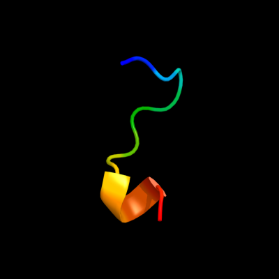
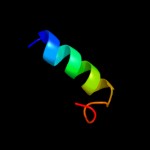
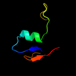
PDB 1vko chain A domain 1
Region: 15 - 27
Aligned: 13
Modelled: 13
Confidence: 28.0%
Identity: 54%
Fold: NAD(P)-binding Rossmann-fold domains
Superfamily: NAD(P)-binding Rossmann-fold domains
Family: Glyceraldehyde-3-phosphate dehydrogenase-like, N-terminal domain
Phyre2
| 2 |
|
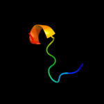
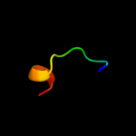
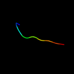
PDB 1p1j chain A domain 1
Region: 15 - 27
Aligned: 13
Modelled: 13
Confidence: 20.3%
Identity: 54%
Fold: NAD(P)-binding Rossmann-fold domains
Superfamily: NAD(P)-binding Rossmann-fold domains
Family: Glyceraldehyde-3-phosphate dehydrogenase-like, N-terminal domain
Phyre2
| 3 |
|
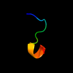
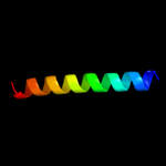
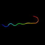
PDB 1vko chain A
Region: 15 - 27
Aligned: 13
Modelled: 13
Confidence: 18.4%
Identity: 54%
PDB header:isomerase
Chain: A: PDB Molecule:inositol-3-phosphate synthase;
PDBTitle: crystal structure of inositol-3-phosphate synthase (ce21227) from2 caenorhabditis elegans at 2.30 a resolution
Phyre2
| 4 |
|
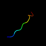
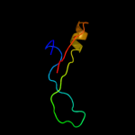
PDB 1pv0 chain A
Region: 58 - 77
Aligned: 20
Modelled: 20
Confidence: 12.8%
Identity: 30%
Fold: Long alpha-hairpin
Superfamily: Sporulation inhibitor Sda
Family: Sporulation inhibitor Sda
Phyre2
| 5 |
|
PDB 1p1h chain D
Region: 15 - 27
Aligned: 13
Modelled: 13
Confidence: 11.8%
Identity: 54%
PDB header:isomerase
Chain: D: PDB Molecule:inositol-3-phosphate synthase;
PDBTitle: crystal structure of the 1l-myo-inositol/nad+ complex
Phyre2
| 6 |
|
PDB 3bj4 chain B
Region: 37 - 63
Aligned: 27
Modelled: 27
Confidence: 11.6%
Identity: 37%
PDB header:signaling protein
Chain: B: PDB Molecule:potassium voltage-gated channel subfamily kqt
PDBTitle: the kcnq1 (kv7.1) c-terminus, a multi-tiered scaffold for2 subunit assembly and protein interaction
Phyre2
| 7 |
|
PDB 2j44 chain A domain 2
Region: 28 - 37
Aligned: 10
Modelled: 10
Confidence: 11.2%
Identity: 30%
Fold: Prealbumin-like
Superfamily: Starch-binding domain-like
Family: PUD-like
Phyre2
| 8 |
|
PDB 2lfe chain A
Region: 13 - 52
Aligned: 38
Modelled: 40
Confidence: 11.2%
Identity: 34%
PDB header:ligase
Chain: A: PDB Molecule:e3 ubiquitin-protein ligase hecw2;
PDBTitle: solution nmr structure of n-terminal domain of human e3 ubiquitin-2 protein ligase hecw2, northeast structural genomics consortium (nesg)3 target ht6306a
Phyre2
| 9 |
|
PDB 2le2 chain B
Region: 21 - 29
Aligned: 9
Modelled: 9
Confidence: 9.0%
Identity: 78%
PDB header:hydrolase inhibitor
Chain: B: PDB Molecule:p56;
PDBTitle: novel dimeric structure of phage phi29-encoded protein p56: insights2 into uracil-dna glycosylase inhibition
Phyre2
| 10 |
|
PDB 2j43 chain A domain 2
Region: 28 - 38
Aligned: 11
Modelled: 11
Confidence: 7.4%
Identity: 27%
Fold: Prealbumin-like
Superfamily: Starch-binding domain-like
Family: PUD-like
Phyre2
| 11 |
|
PDB 2qbx chain B
Region: 10 - 54
Aligned: 38
Modelled: 45
Confidence: 6.6%
Identity: 37%
PDB header:signaling protein
Chain: B: PDB Molecule:ephrin type-b receptor 2;
PDBTitle: ephb2/snew antagonistic peptide complex
Phyre2