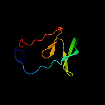
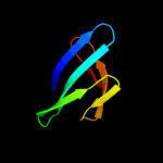
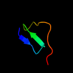
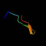
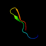
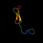
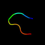
PDB header:lipoprotein Chain: A: PDB Molecule:pilp pilot protein; PDBTitle: the solution structure of a domain from the neisseria2 meningitidis pilp pilot protein.
Confidence and coverage
Confidence:
96.7%
Coverage:
56%
75 residues ( 56% of your sequence) have been modelled with 96.7% confidence by the single highest scoring template.
You may wish to submit your sequence to Phyrealarm. This will automatically scan your sequence every week for new potential templates as they appear in the Phyre2 library.
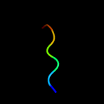
Region: 17 - 92 Aligned: 75 Modelled: 76 Confidence: 96.7% Identity: 21% PDB header:lipoprotein Chain: A: PDB Molecule:pilp pilot protein; PDBTitle: the solution structure of a domain from the neisseria2 meningitidis pilp pilot protein.
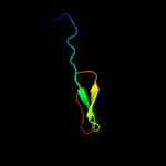
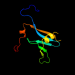
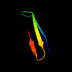
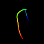
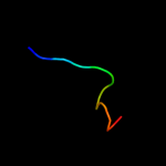
Region: 39 - 87 Aligned: 47 Modelled: 49 Confidence: 83.8% Identity: 19% PDB header:protein transport Chain: C: PDB Molecule:type 2 secretion system, gspc; PDBTitle: the crystal structure of enterotoxigenic escherichia coli gspc-gspd2 complex from the type ii secretion system
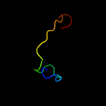
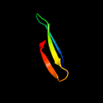
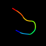
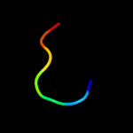
Region: 56 - 84 Aligned: 28 Modelled: 29 Confidence: 12.4% Identity: 32% PDB header:ribosomal protein Chain: C: PDB Molecule:ribosomal protein s2-related protein; PDBTitle: the crystal structure of dr2241 from deinococcus2 radiodurans at 1.9 a resolution reveals a multi-domain3 protein with structural similarity to chelatases but also4 with two additional novel domains
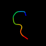
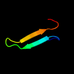
Region: 70 - 76 Aligned: 7 Modelled: 7 Confidence: 7.8% Identity: 29% PDB header:signaling protein/transcription Chain: A: PDB Molecule:e3 ubiquitin-protein ligase smurf1; PDBTitle: structure of the first ww domain of human smurf1 in complex with a di-2 phosphorylated human smad1 derived peptide