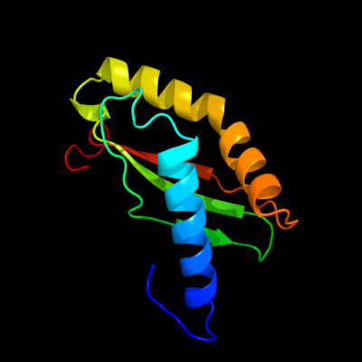
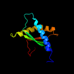
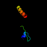
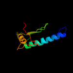
1 d1kkga_
100.0
100
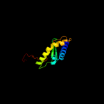
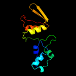
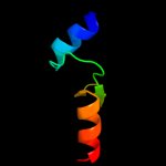
Fold: Alpha-lytic protease prodomain-likeSuperfamily: Ribosome-binding factor A, RbfAFamily: Ribosome-binding factor A, RbfA2 d1josa_
100.0
71
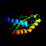
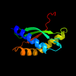
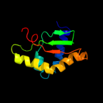
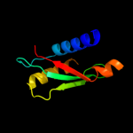
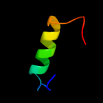
Fold: Alpha-lytic protease prodomain-likeSuperfamily: Ribosome-binding factor A, RbfAFamily: Ribosome-binding factor A, RbfA3 c2kzfA_
100.0
36
PDB header: ribosomal proteinChain: A: PDB Molecule: ribosome-binding factor a;PDBTitle: solution nmr structure of the thermotoga maritima protein tm0855 a2 putative ribosome binding factor a
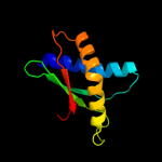
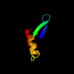
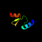
4 d2e7ga1
100.0
18
Fold: Alpha-lytic protease prodomain-likeSuperfamily: Ribosome-binding factor A, RbfAFamily: Ribosome-binding factor A, RbfA5 d1pa4a_
100.0
20
Fold: Alpha-lytic protease prodomain-likeSuperfamily: Ribosome-binding factor A, RbfAFamily: Ribosome-binding factor A, RbfA6 d2dyja1
100.0
27
Fold: Alpha-lytic protease prodomain-likeSuperfamily: Ribosome-binding factor A, RbfAFamily: Ribosome-binding factor A, RbfA7 c2qsiB_
12.4
10
PDB header: structural genomics, unknown functionChain: B: PDB Molecule: putative hydrogenase expression/formation protein hupg;PDBTitle: crystal structure of putative hydrogenase expression/formation protein2 hupg from rhodopseudomonas palustris cga009
8 c3ereD_
9.6
17
PDB header: dna binding protein/dnaChain: D: PDB Molecule: arginine repressor;PDBTitle: crystal structure of the arginine repressor protein from mycobacterium2 tuberculosis in complex with the dna operator
9 d1lr0a_
9.2
14
Fold: TolA/TonB C-terminal domainSuperfamily: TolA/TonB C-terminal domainFamily: TolA10 c3kh0A_
9.2
20
PDB header: signaling proteinChain: A: PDB Molecule: ral guanine nucleotide dissociation stimulator;PDBTitle: crystal structure of the ras-association (ra) domain of2 ralgds
11 d1jmta_
7.6
19
Fold: Ferredoxin-likeSuperfamily: RNA-binding domain, RBDFamily: Splicing factor U2AF subunits12 c3c1sA_
7.4
15
PDB header: oxidoreductaseChain: A: PDB Molecule: glutaredoxin-1;PDBTitle: crystal structure of grx1 in glutathionylated form
13 d2rgfa_
7.1
20
Fold: beta-Grasp (ubiquitin-like)Superfamily: Ubiquitin-likeFamily: Ras-binding domain, RBD14 c2jacA_
7.0
16
PDB header: electron transportChain: A: PDB Molecule: glutaredoxin-1;PDBTitle: glutaredoxin grx1p c30s mutant from yeast
15 d2b3aa1
6.9
20
Fold: beta-Grasp (ubiquitin-like)Superfamily: Ubiquitin-likeFamily: Ras-binding domain, RBD16 d1ug8a_
6.7
20
Fold: IF3-likeSuperfamily: R3H domainFamily: R3H domain17 c3fzaA_
6.6
15
PDB header: oxidoreductaseChain: A: PDB Molecule: glutaredoxin;PDBTitle: crystal structure of poplar glutaredoxin s12 in complex with2 glutathione and beta-mercaptoethanol
18 d1lfda_
6.4
20
Fold: beta-Grasp (ubiquitin-like)Superfamily: Ubiquitin-likeFamily: Ras-binding domain, RBD19 d1l0wa2
6.4
24
Fold: DCoH-likeSuperfamily: GAD domain-likeFamily: GAD domain20 c3ipzA_
6.3
11
PDB header: electron transport, oxidoreductaseChain: A: PDB Molecule: monothiol glutaredoxin-s14, chloroplastic;PDBTitle: crystal structure of arabidopsis monothiol glutaredoxin atgrxcp
21 d1xmka1
not modelled
6.3
15
Fold: DNA/RNA-binding 3-helical bundleSuperfamily: "Winged helix" DNA-binding domainFamily: Z-DNA binding domain22 c2gboB_
not modelled
6.2
17
PDB header: structural genomics, unknown functionChain: B: PDB Molecule: upf0358 protein ef2458;PDBTitle: protein of unknown function ef2458 from enterococcus faecalis
23 d2gboa1
not modelled
6.2
17
Fold: Open three-helical up-and-down bundleSuperfamily: EF2458-likeFamily: EF2458-like24 d1whxa_
not modelled
6.2
16
Fold: Ferredoxin-likeSuperfamily: RNA-binding domain, RBDFamily: Canonical RBD25 c3d5jB_
not modelled
5.8
13
PDB header: oxidoreductaseChain: B: PDB Molecule: glutaredoxin-2, mitochondrial;PDBTitle: structure of yeast grx2-c30s mutant with glutathionyl mixed2 disulfide
26 c3nm7D_
not modelled
5.7
19
PDB header: nucleic acid binding proteinChain: D: PDB Molecule: uncharacterized protein;PDBTitle: crystal structure of borrelia burgdorferi pur-alpha
27 d1hska2
not modelled
5.2
17
Fold: Uridine diphospho-N-Acetylenolpyruvylglucosamine reductase, MurB, C-terminal domainSuperfamily: Uridine diphospho-N-Acetylenolpyruvylglucosamine reductase, MurB, C-terminal domainFamily: Uridine diphospho-N-Acetylenolpyruvylglucosamine reductase, MurB, C-terminal domain28 d1ep7a_
not modelled
5.1
15
Fold: Thioredoxin foldSuperfamily: Thioredoxin-likeFamily: Thioltransferase