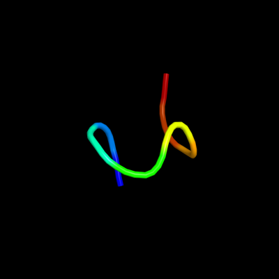
Phyre Home Retrieve Phyre Job Id
Emaill.a.kelley@imperial.ac.uk
DescriptionP75971
DateThu Jan 5 12:16:39 GMT 2012
Unique Job ID22c759e81db252f0

Summary 

Top model
Image coloured by rainbow N → C terminus
Model (left) based on template c2iurD_
Top template information
PDB header:oxidoreductase
Chain: D: PDB Molecule:aromatic amine dehydrogenase beta subunit;
PDBTitle: crystal structure of n-quinol form of aromatic amine2 dehydrogenase (aadh) from alcaligenes faecalis, form a3 cocrystal
Confidence and coverage
Confidence: 9.9% Coverage: 13%
13 residues ( 13% of your sequence) have been modelled with 9.9% confidence by the single highest scoring template.
You may wish to submit your sequence to Phyrealarm. This will automatically scan your sequence every week for new potential templates as they appear in the Phyre2 library.
Please note: You must be registered and logged in to use Phyrealarm.
Warning: 61% of your sequence is predicted disordered. Disordered regions cannot be meaningfully predicted.
3D viewing
Interactive 3D view in Jmol

Sequence analysis 

Secondary structure and disorder prediction 

   1........10.........20.........30.........40.........50.........60
Sequence  MVNAAQRTRVKVEADNRPSVDTHPPGVQPSPGTGGTRHHNFMLCVVLAVPVFSLVLSGTA
Secondary structure 


SS confidence 


Disorder  ???????


?????????????

?????????


???????????
Disorder confidence 


   .........70.........80.........90.........100...
Sequence  LFTKQRRVSPDDGLITRPILIAVATGALLCFVEKLTDRAGSIC
Secondary structure 


SS confidence 


Disorder  ??????????????
??


???????
Disorder confidence 


Confidence Key
High(9)                    Low (0)
?Disordered
Alpha helix
Beta strand

Domain analysis 

Hover over an aligned region to see model and summary info

Please note, only up to the top 20 hits are modelled to reduce computer load

RankAligned region

PDB 2iur chain D

3D model

Region: 19 - 31
Aligned: 13
Modelled: 13
Confidence: 9.9%
Identity: 54%
PDB header:oxidoreductase
Chain: D: PDB Molecule:aromatic amine dehydrogenase beta subunit;
PDBTitle: crystal structure of n-quinol form of aromatic amine2 dehydrogenase (aadh) from alcaligenes faecalis, form a3 cocrystal

Phyre2

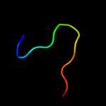
PDB 2awi chain A domain 1

3D model

Region: 11 - 18
Aligned: 8
Modelled: 8
Confidence: 9.0%
Identity: 88%
Fold: lambda repressor-like DNA-binding domains
Superfamily: lambda repressor-like DNA-binding domains
Family: PrgX N-terminal domain-like

Phyre2

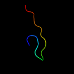
PDB 1prt chain C domain 2

3D model

Region: 54 - 65
Aligned: 12
Modelled: 12
Confidence: 7.4%
Identity: 50%
Fold: C-type lectin-like
Superfamily: C-type lectin-like
Family: Aerolysin/Pertussis toxin (APT) domain

Phyre2

PDB 1mda chain L

3D model

Region: 19 - 36
Aligned: 18
Modelled: 18
Confidence: 6.2%
Identity: 33%
Fold: Methylamine dehydrogenase, L chain
Superfamily: Methylamine dehydrogenase, L chain
Family: Methylamine dehydrogenase, L chain

Phyre2

PDB 3c75 chain L

3D model

Region: 19 - 36
Aligned: 18
Modelled: 18
Confidence: 5.5%
Identity: 22%
PDB header:oxidoreductase
Chain: L: PDB Molecule:methylamine dehydrogenase light chain;
PDBTitle: paracoccus versutus methylamine dehydrogenase in complex2 with amicyanin

Phyre2

PDB 1w36 chain B domain 1

3D model

Region: 27 - 84
Aligned: 50
Modelled: 58
Confidence: 5.5%
Identity: 26%
Fold: P-loop containing nucleoside triphosphate hydrolases
Superfamily: P-loop containing nucleoside triphosphate hydrolases
Family: Tandem AAA-ATPase domain

Phyre2

PDB 2bbk chain L

3D model

Region: 19 - 36
Aligned: 18
Modelled: 18
Confidence: 5.5%
Identity: 22%
Fold: Methylamine dehydrogenase, L chain
Superfamily: Methylamine dehydrogenase, L chain
Family: Methylamine dehydrogenase, L chain

Phyre2
1

c2iurD_
2

d2awia1
3

d1prtc2
4

d1mdal_
5

c3c75L_
6

d1w36b1
7

d2bbkl_


Detailed template information 

#
Template Alignment Coverage3D Model Confidence
% i.d. Template Information
1c2iurD_


9.9 54 PDB header:oxidoreductase
Chain: D: PDB Molecule:aromatic amine dehydrogenase beta subunit;
PDBTitle: crystal structure of n-quinol form of aromatic amine2 dehydrogenase (aadh) from alcaligenes faecalis, form a3 cocrystal
2d2awia1


9.0 88 Fold:lambda repressor-like DNA-binding domains
Superfamily:lambda repressor-like DNA-binding domains
Family:PrgX N-terminal domain-like
3d1prtc2


7.4 50 Fold:C-type lectin-like
Superfamily:C-type lectin-like
Family:Aerolysin/Pertussis toxin (APT) domain
4d1mdal_


6.2 33 Fold:Methylamine dehydrogenase, L chain
Superfamily:Methylamine dehydrogenase, L chain
Family:Methylamine dehydrogenase, L chain
5c3c75L_


5.5 22 PDB header:oxidoreductase
Chain: L: PDB Molecule:methylamine dehydrogenase light chain;
PDBTitle: paracoccus versutus methylamine dehydrogenase in complex2 with amicyanin
6d1w36b1


5.5 26 Fold:P-loop containing nucleoside triphosphate hydrolases
Superfamily:P-loop containing nucleoside triphosphate hydrolases
Family:Tandem AAA-ATPase domain
7d2bbkl_


5.5 22 Fold:Methylamine dehydrogenase, L chain
Superfamily:Methylamine dehydrogenase, L chain
Family:Methylamine dehydrogenase, L chain

Binding site prediction 

Due to computational demand, binding site predictions are not run for batch jobs

If you want to predict binding sites, please manually submit your model of choice to 3DLigandSite


Phyre is for academic use only

Please cite: Protein structure prediction on the web: a case study using the Phyre server
Kelley LA and Sternberg MJE. Nature Protocols 4, 363 - 371 (2009) [pdf] [Import into BibTeX]
 
If you use the binding site predictions from 3DLigandSite, please also cite:
3DLigandSite: predicting ligand-binding sites using similar structures.
Wass MN, Kelley LA and Sternberg MJ Nucleic Acids Research 38, W469-73 (2010) [PubMed]
 
© Structural Bioinformatics Group
Imperial College London
Lawrence Kelley, Benjamin Jefferys
Disclaimer
Terms and Conditions
Component software
Template detection: HHpred 1.51
Secondary structure prediction: Psi-pred 2.5
Disorder prediction: Disopred 2.4
Transmembrane prediction: Memsat_SVM
Multi-template modelling and ab initio: Poing 1.0