| 1 |
|
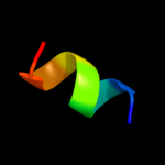
PDB 1m2z chain E
Region: 16 - 24
Aligned: 9
Modelled: 9
Confidence: 14.2%
Identity: 33%
PDB header:hormone/hormone activator
Chain: E: PDB Molecule:nuclear receptor coactivator 2;
PDBTitle: crystal structure of a dimer complex of the human2 glucocorticoid receptor ligand-binding domain bound to3 dexamethasone and a tif2 coactivator motif
Phyre2
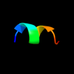
| 2 |
|
PDB 3gn8 chain C
Region: 16 - 24
Aligned: 9
Modelled: 9
Confidence: 11.4%
Identity: 33%
PDB header:hormone/hormone activator
Chain: C: PDB Molecule:nuclear receptor coactivator 2;
PDBTitle: x-ray crystal structure of ancgr2 in complex with2 dexamethasone
Phyre2
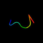
| 3 |
|
PDB 3gn8 chain E
Region: 16 - 24
Aligned: 9
Modelled: 9
Confidence: 10.7%
Identity: 33%
PDB header:hormone/hormone activator
Chain: E: PDB Molecule:nuclear receptor coactivator 2;
PDBTitle: x-ray crystal structure of ancgr2 in complex with2 dexamethasone
Phyre2
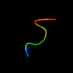
| 4 |
|
PDB 1m2z chain B
Region: 16 - 24
Aligned: 9
Modelled: 9
Confidence: 9.6%
Identity: 33%
PDB header:hormone/hormone activator
Chain: B: PDB Molecule:nuclear receptor coactivator 2;
PDBTitle: crystal structure of a dimer complex of the human2 glucocorticoid receptor ligand-binding domain bound to3 dexamethasone and a tif2 coactivator motif
Phyre2
| 5 |
|
PDB 2xok chain G
Region: 72 - 79
Aligned: 8
Modelled: 8
Confidence: 8.9%
Identity: 38%
PDB header:hydrolase
Chain: G: PDB Molecule:atp synthase subunit gamma, mitochondrial;
PDBTitle: refined structure of yeast f1c10 atpase complex to 3 a2 resolution
Phyre2
| 6 |
|
PDB 2qe7 chain G
Region: 72 - 79
Aligned: 8
Modelled: 8
Confidence: 6.5%
Identity: 25%
PDB header:hydrolase
Chain: G: PDB Molecule:atp synthase subunit gamma;
PDBTitle: crystal structure of the f1-atpase from the thermoalkaliphilic2 bacterium bacillus sp. ta2.a1
Phyre2
| 7 |
|
PDB 2jdi chain G domain 1
Region: 72 - 79
Aligned: 8
Modelled: 8
Confidence: 6.4%
Identity: 38%
Fold: Pyruvate kinase C-terminal domain-like
Superfamily: ATP synthase (F1-ATPase), gamma subunit
Family: ATP synthase (F1-ATPase), gamma subunit
Phyre2
| 8 |
|
PDB 1fs0 chain G
Region: 72 - 79
Aligned: 8
Modelled: 8
Confidence: 6.2%
Identity: 63%
Fold: Pyruvate kinase C-terminal domain-like
Superfamily: ATP synthase (F1-ATPase), gamma subunit
Family: ATP synthase (F1-ATPase), gamma subunit
Phyre2
| 9 |
|
PDB 1u7g chain A
Region: 7 - 85
Aligned: 79
Modelled: 79
Confidence: 6.2%
Identity: 14%
Fold: Ammonium transporter
Superfamily: Ammonium transporter
Family: Ammonium transporter
Phyre2
| 10 |
|
PDB 1sxd chain A
Region: 75 - 86
Aligned: 12
Modelled: 12
Confidence: 5.7%
Identity: 8%
Fold: SAM domain-like
Superfamily: SAM/Pointed domain
Family: Pointed domain
Phyre2