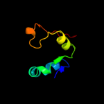
1 c3au9A_
37.0
18
PDB header: isomerase/isomerase inhibitorChain: A: PDB Molecule: 1-deoxy-d-xylulose 5-phosphate reductoisomerase;PDBTitle: crystal structure of the quaternary complex-1 of an isomerase
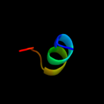
2 d2ijra1
22.7
24
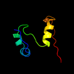
Fold: Api92-likeSuperfamily: Api92-likeFamily: Api92-like3 d1ei5a1
19.7
30
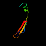
Fold: Streptavidin-likeSuperfamily: D-aminopeptidase, middle and C-terminal domainsFamily: D-aminopeptidase, middle and C-terminal domains4 d1zy9a1
18.5
33
Fold: SupersandwichSuperfamily: Galactose mutarotase-likeFamily: YicI N-terminal domain-like5 c2jrbA_
17.0
43
PDB header: rna binding proteinChain: A: PDB Molecule: orf 1 protein;PDBTitle: c-terminal domain of orf1p from mouse line-1
6 d1smpi_
15.1
23
Fold: Streptavidin-likeSuperfamily: beta-Barrel protease inhibitorsFamily: Metalloprotease inhibitor7 c1r0lD_
12.5
21
PDB header: oxidoreductaseChain: D: PDB Molecule: 1-deoxy-d-xylulose 5-phosphate reductoisomerase;PDBTitle: 1-deoxy-d-xylulose 5-phosphate reductoisomerase from2 zymomonas mobilis in complex with nadph
8 c1k5hB_
9.3
20
PDB header: oxidoreductaseChain: B: PDB Molecule: 1-deoxy-d-xylulose-5-phosphate reductoisomerase;PDBTitle: 1-deoxy-d-xylulose-5-phosphate reductoisomerase
9 d2ctda1
8.6
31
Fold: beta-beta-alpha zinc fingersSuperfamily: beta-beta-alpha zinc fingersFamily: Classic zinc finger, C2H210 c2jcyA_
8.4
19
PDB header: oxidoreductaseChain: A: PDB Molecule: 1-deoxy-d-xylulose 5-phosphate reductoisomerase;PDBTitle: x-ray structure of mutant 1-deoxy-d-xylulose 5-phosphate2 reductoisomerase, dxr, rv2870c, from mycobacterium3 tuberculosis
11 d1ggla_
7.4
15
Fold: LipocalinsSuperfamily: LipocalinsFamily: Fatty acid binding protein-like12 d1uowa_
7.3
20
Fold: C2 domain-likeSuperfamily: C2 domain (Calcium/lipid-binding domain, CaLB)Family: Synaptotagmin-like (S variant)13 c2x3bB_
7.0
38
PDB header: hydrolaseChain: B: PDB Molecule: toxic extracellular endopeptidase;PDBTitle: asap1 inactive mutant e294a, an extracellular toxic zinc2 metalloendopeptidase
14 c1a6cA_
6.8
15
PDB header: virusChain: A: PDB Molecule: tobacco ringspot virus capsid protein;PDBTitle: structure of tobacco ringspot virus
15 c3a14B_
6.6
19
PDB header: oxidoreductaseChain: B: PDB Molecule: 1-deoxy-d-xylulose 5-phosphate reductoisomerase;PDBTitle: crystal structure of dxr from thermotoga maritima, in complex with2 nadph
16 d1j3na1
6.5
15
Fold: Thiolase-likeSuperfamily: Thiolase-likeFamily: Thiolase-related17 c3monF_
6.3
42
PDB header: sweet-tasting proteinChain: F: PDB Molecule: monellin;PDBTitle: crystal structures of two intensely sweet proteins
18 d1r0ka3
6.2
17
Fold: FwdE/GAPDH domain-likeSuperfamily: Glyceraldehyde-3-phosphate dehydrogenase-like, C-terminal domainFamily: Dihydrodipicolinate reductase-like19 d1bu2a2
6.2
50
Fold: Cyclin-likeSuperfamily: Cyclin-likeFamily: Cyclin20 d1eg1a_
6.1
22
Fold: Concanavalin A-like lectins/glucanasesSuperfamily: Concanavalin A-like lectins/glucanasesFamily: Glycosyl hydrolase family 7 catalytic core21 c3hmhA_
not modelled
5.5
25
PDB header: transcriptionChain: A: PDB Molecule: transcription initiation factor tfiid 210 kda subunit;PDBTitle: crystal structure of the second bromodomain of human tbp-associated2 factor rna polymerase 1-like (taf1l)
22 c3p19A_
not modelled
5.3
40
PDB header: oxidoreductaseChain: A: PDB Molecule: putative blue fluorescent protein;PDBTitle: improved nadph-dependent blue fluorescent protein
23 d1q0qa3
not modelled
5.1
18
Fold: FwdE/GAPDH domain-likeSuperfamily: Glyceraldehyde-3-phosphate dehydrogenase-like, C-terminal domainFamily: Dihydrodipicolinate reductase-like24 c2eghA_
not modelled
5.1
18
PDB header: oxidoreductaseChain: A: PDB Molecule: 1-deoxy-d-xylulose 5-phosphate reductoisomerase;PDBTitle: crystal structure of 1-deoxy-d-xylulose 5-phosphate reductoisomerase2 complexed with a magnesium ion, nadph and fosmidomycin