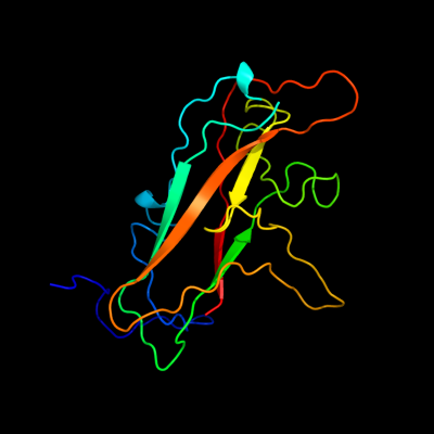
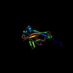
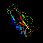
| 1 |
|
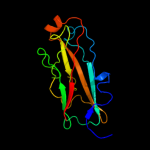
PDB 2jty chain A
Region: 26 - 200
Aligned: 155
Modelled: 175
Confidence: 99.9%
Identity: 23%
PDB header:structural protein
Chain: A: PDB Molecule:type-1 fimbrial protein, a chain;
PDBTitle: self-complemented variant of fima, the main subunit of type 1 pilus
Phyre2
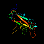
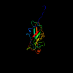
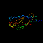
| 2 |
|
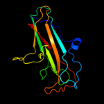
PDB 3jwn chain K
Region: 31 - 200
Aligned: 150
Modelled: 165
Confidence: 99.9%
Identity: 21%
PDB header:protein binding/cell adhesion
Chain: K: PDB Molecule:protein fimf;
PDBTitle: complex of fimc, fimf, fimg and fimh
Phyre2
| 3 |
|
PDB 3jwn chain L
Region: 31 - 200
Aligned: 150
Modelled: 170
Confidence: 99.9%
Identity: 21%
PDB header:protein binding/cell adhesion
Chain: L: PDB Molecule:protein fimf;
PDBTitle: complex of fimc, fimf, fimg and fimh
Phyre2
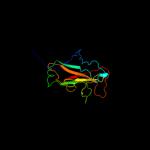
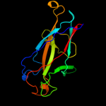
| 4 |
|
PDB 3jwn chain E
Region: 31 - 200
Aligned: 150
Modelled: 164
Confidence: 99.9%
Identity: 21%
PDB header:protein binding/cell adhesion
Chain: E: PDB Molecule:protein fimf;
PDBTitle: complex of fimc, fimf, fimg and fimh
Phyre2
| 5 |
|
PDB 3jwn chain F
Region: 31 - 200
Aligned: 150
Modelled: 170
Confidence: 99.9%
Identity: 21%
PDB header:protein binding/cell adhesion
Chain: F: PDB Molecule:protein fimf;
PDBTitle: complex of fimc, fimf, fimg and fimh
Phyre2
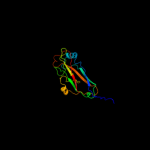
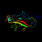
| 6 |
|
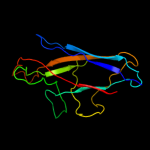
PDB 2jmr chain A
Region: 31 - 200
Aligned: 150
Modelled: 160
Confidence: 99.9%
Identity: 21%
PDB header:cell adhesion
Chain: A: PDB Molecule:fimf;
PDBTitle: nmr structure of the e. coli type 1 pilus subunit fimf
Phyre2
| 7 |
|
PDB 2uy6 chain B domain 1
Region: 34 - 200
Aligned: 147
Modelled: 155
Confidence: 99.9%
Identity: 21%
Fold: Common fold of diphtheria toxin/transcription factors/cytochrome f
Superfamily: Bacterial adhesins
Family: Pilus subunits
Phyre2
| 8 |
|
PDB 2j2z chain B domain 1
Region: 35 - 200
Aligned: 146
Modelled: 156
Confidence: 99.8%
Identity: 12%
Fold: Common fold of diphtheria toxin/transcription factors/cytochrome f
Superfamily: Bacterial adhesins
Family: Pilus subunits
Phyre2
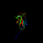
| 9 |
|
PDB 1pdk chain B
Region: 41 - 200
Aligned: 143
Modelled: 150
Confidence: 99.8%
Identity: 15%
Fold: Common fold of diphtheria toxin/transcription factors/cytochrome f
Superfamily: Bacterial adhesins
Family: Pilus subunits
Phyre2
| 10 |
|
PDB 3bfw chain A
Region: 43 - 201
Aligned: 129
Modelled: 149
Confidence: 99.8%
Identity: 18%
PDB header:structural protein/structural protein
Chain: A: PDB Molecule:protein fimg;
PDBTitle: crystal structure of truncated fimg (fimgt) in complex with the donor2 strand peptide of fimf (dsf)
Phyre2
| 11 |
|
PDB 3bwu chain F
Region: 43 - 200
Aligned: 127
Modelled: 143
Confidence: 99.7%
Identity: 24%
PDB header:chaperone, structural, membrane protein
Chain: F: PDB Molecule:protein fimf;
PDBTitle: crystal structure of the ternary complex of fimd (n-terminal domain,2 fimdn) with fimc and the n-terminally truncated pilus subunit fimf3 (fimft)
Phyre2
| 12 |
|
PDB 2w07 chain B
Region: 40 - 201
Aligned: 121
Modelled: 137
Confidence: 99.7%
Identity: 23%
PDB header:cell adhesion
Chain: B: PDB Molecule:minor pilin subunit papf;
PDBTitle: structural determinants of polymerization reactivity of the2 p pilus adaptor subunit papf
Phyre2
| 13 |
|
PDB 1ze3 chain H domain 1
Region: 44 - 201
Aligned: 121
Modelled: 146
Confidence: 99.6%
Identity: 21%
Fold: Common fold of diphtheria toxin/transcription factors/cytochrome f
Superfamily: Bacterial adhesins
Family: Pilus subunits
Phyre2
| 14 |
|
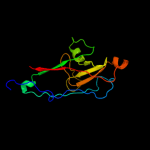
PDB 1klf chain P
Region: 40 - 200
Aligned: 124
Modelled: 142
Confidence: 99.5%
Identity: 22%
PDB header:chaperone/adhesin complex
Chain: P: PDB Molecule:fimh protein;
PDBTitle: fimh adhesin-fimc chaperone complex with d-mannose
Phyre2
| 15 |
|
PDB 1n12 chain A
Region: 44 - 201
Aligned: 136
Modelled: 150
Confidence: 99.5%
Identity: 21%
Fold: Common fold of diphtheria toxin/transcription factors/cytochrome f
Superfamily: Bacterial adhesins
Family: Pilus subunits
Phyre2
| 16 |
|
PDB 2omz chain A domain 1
Region: 31 - 44
Aligned: 14
Modelled: 14
Confidence: 9.1%
Identity: 21%
Fold: Immunoglobulin-like beta-sandwich
Superfamily: E set domains
Family: Internalin Ig-like domain
Phyre2
| 17 |
|
PDB 1h6t chain A domain 1
Region: 31 - 44
Aligned: 14
Modelled: 14
Confidence: 8.6%
Identity: 7%
Fold: Immunoglobulin-like beta-sandwich
Superfamily: E set domains
Family: Internalin Ig-like domain
Phyre2