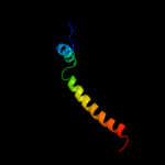
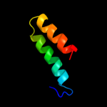
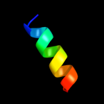
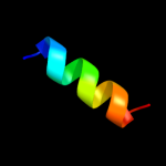
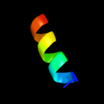
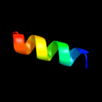
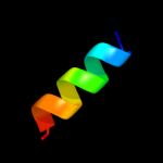
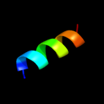
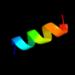
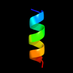
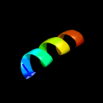
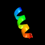
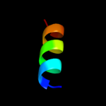
1 c2l16A_
99.8
27
PDB header: protein transportChain: A: PDB Molecule: sec-independent protein translocase protein tatad;PDBTitle: solution structure of bacillus subtilits tatad protein in dpc micelles
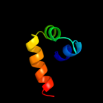
2 d2axtj1
49.7
31
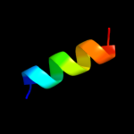
Fold: Single transmembrane helixSuperfamily: Photosystem II reaction center protein J, PsbJFamily: PsbJ-like3 d1ohua_
45.3
13
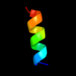
Fold: Toxins' membrane translocation domainsSuperfamily: Bcl-2 inhibitors of programmed cell deathFamily: Bcl-2 inhibitors of programmed cell death4 d2axti1
37.6
31
Fold: Single transmembrane helixSuperfamily: Photosystem II reaction center protein I, PsbIFamily: PsbI-like5 d1bxla_
30.0
18
Fold: Toxins' membrane translocation domainsSuperfamily: Bcl-2 inhibitors of programmed cell deathFamily: Bcl-2 inhibitors of programmed cell death6 d2cqqa1
29.8
15
Fold: DNA/RNA-binding 3-helical bundleSuperfamily: Homeodomain-likeFamily: Myb/SANT domain7 c3prrL_
29.5
29
PDB header: photosynthesisChain: L: PDB Molecule: photosystem ii reaction center protein l;PDBTitle: crystal structure of cyanobacterial photosystem ii in complex with2 terbutryn (part 2 of 2). this file contains second monomer of psii3 dimer
8 c1s5lL_
29.5
29
PDB header: photosynthesisChain: L: PDB Molecule: photosystem ii reaction center l protein;PDBTitle: architecture of the photosynthetic oxygen evolving center
9 c3bz1L_
29.5
29
PDB header: electron transportChain: L: PDB Molecule: photosystem ii reaction center protein l;PDBTitle: crystal structure of cyanobacterial photosystem ii (part 12 of 2). this file contains first monomer of psii dimer
10 c3bz2L_
29.5
29
PDB header: electron transportChain: L: PDB Molecule: photosystem ii reaction center protein l;PDBTitle: crystal structure of cyanobacterial photosystem ii (part 22 of 2). this file contains second monomer of psii dimer
11 c3prqL_
29.5
29
PDB header: photosynthesisChain: L: PDB Molecule: photosystem ii reaction center protein l;PDBTitle: crystal structure of cyanobacterial photosystem ii in complex with2 terbutryn (part 1 of 2). this file contains first monomer of psii3 dimer
12 c3arcL_
29.5
29
PDB header: electron transport, photosynthesisChain: L: PDB Molecule: photosystem ii reaction center protein l;PDBTitle: crystal structure of oxygen-evolving photosystem ii at 1.9 angstrom2 resolution
13 d2axtl1
29.5
29
Fold: Single transmembrane helixSuperfamily: Photosystem II reaction center protein L, PsbLFamily: PsbL-like14 c2axtl_
29.5
29
PDB header: electron transportChain: L: PDB Molecule: photosystem ii reaction center l protein;PDBTitle: crystal structure of photosystem ii from thermosynechococcus elongatus
15 c1s5ll_
29.5
29
PDB header: photosynthesisChain: L: PDB Molecule: photosystem ii reaction center l protein;PDBTitle: architecture of the photosynthetic oxygen evolving center
16 c3a0bL_
29.5
29
PDB header: electron transportChain: L: PDB Molecule: photosystem ii reaction center protein l;PDBTitle: crystal structure of br-substituted photosystem ii complex
17 c3a0hL_
29.5
29
PDB header: electron transportChain: L: PDB Molecule: photosystem ii reaction center protein l;PDBTitle: crystal structure of i-substituted photosystem ii complex
18 c3kziL_
29.5
29
PDB header: electron transportChain: L: PDB Molecule: photosystem ii reaction center protein l;PDBTitle: crystal structure of monomeric form of cyanobacterial photosystem ii
19 c2axtL_
29.5
29
PDB header: electron transportChain: L: PDB Molecule: photosystem ii reaction center l protein;PDBTitle: crystal structure of photosystem ii from thermosynechococcus elongatus
20 c3a0hl_
29.5
29
PDB header: electron transportChain: L: PDB Molecule: photosystem ii reaction center protein l;PDBTitle: crystal structure of i-substituted photosystem ii complex
21 c3a0bl_
not modelled
29.5
29
PDB header: electron transportChain: L: PDB Molecule: photosystem ii reaction center protein l;PDBTitle: crystal structure of br-substituted photosystem ii complex
22 d1pq1a_
not modelled
24.4
16
Fold: Toxins' membrane translocation domainsSuperfamily: Bcl-2 inhibitors of programmed cell deathFamily: Bcl-2 inhibitors of programmed cell death23 c3arcl_
not modelled
23.0
29
PDB header: electron transport, photosynthesisChain: L: PDB Molecule: photosystem ii reaction center protein l;PDBTitle: crystal structure of oxygen-evolving photosystem ii at 1.9 angstrom2 resolution
24 c2bzwB_
not modelled
22.2
15
PDB header: transcriptionChain: B: PDB Molecule: bcl2-antagonist of cell death;PDBTitle: the crystal structure of bcl-xl in complex with full-length2 bad
25 c3qbrA_
not modelled
21.2
16
PDB header: apoptosisChain: A: PDB Molecule: sjchgc06286 protein;PDBTitle: bakbh3 in complex with sja
26 d1zy3a1
not modelled
20.1
11
Fold: Toxins' membrane translocation domainsSuperfamily: Bcl-2 inhibitors of programmed cell deathFamily: Bcl-2 inhibitors of programmed cell death27 d2jm6b1
not modelled
16.7
15
Fold: Toxins' membrane translocation domainsSuperfamily: Bcl-2 inhibitors of programmed cell deathFamily: Bcl-2 inhibitors of programmed cell death28 c2voyH_
not modelled
16.1
20
PDB header: hydrolaseChain: H: PDB Molecule: sarcoplasmic/endoplasmic reticulum calciumPDBTitle: cryoem model of copa, the copper transporting atpase from2 archaeoglobus fulgidus
29 d1ysga1
not modelled
14.8
16
Fold: Toxins' membrane translocation domainsSuperfamily: Bcl-2 inhibitors of programmed cell deathFamily: Bcl-2 inhibitors of programmed cell death30 c1yceD_
not modelled
14.4
25
PDB header: membrane proteinChain: D: PDB Molecule: subunit c;PDBTitle: structure of the rotor ring of f-type na+-atpase from ilyobacter2 tartaricus
31 c2nogA_
not modelled
14.1
7
PDB header: dna binding proteinChain: A: PDB Molecule: iswi protein;PDBTitle: sant domain structure of xenopus remodeling factor iswi
32 c1vf5U_
not modelled
12.5
45
PDB header: photosynthesisChain: U: PDB Molecule: protein pet n;PDBTitle: crystal structure of cytochrome b6f complex from m.laminosus
33 c1vf5H_
not modelled
12.5
45
PDB header: photosynthesisChain: H: PDB Molecule: protein pet n;PDBTitle: crystal structure of cytochrome b6f complex from m.laminosus
34 c1g5jB_
not modelled
12.2
23
PDB header: apoptosisChain: B: PDB Molecule: bad protein;PDBTitle: complex of bcl-xl with peptide from bad
35 c2rocB_
not modelled
11.8
17
PDB header: apoptosisChain: B: PDB Molecule: bcl-2-binding component 3;PDBTitle: solution structure of mcl-1 complexed with puma
36 c2yv6A_
not modelled
11.0
16
PDB header: apoptosisChain: A: PDB Molecule: bcl-2 homologous antagonist/killer;PDBTitle: crystal structure of human bcl-2 family protein bak
37 c2zjsE_
not modelled
10.9
21
PDB header: protein transport/immune systemChain: E: PDB Molecule: preprotein translocase sece subunit;PDBTitle: crystal structure of secye translocon from thermus thermophilus with a2 fab fragment
38 d1o0la_
not modelled
10.6
11
Fold: Toxins' membrane translocation domainsSuperfamily: Bcl-2 inhibitors of programmed cell deathFamily: Bcl-2 inhibitors of programmed cell death39 d1g5ma_
not modelled
10.6
11
Fold: Toxins' membrane translocation domainsSuperfamily: Bcl-2 inhibitors of programmed cell deathFamily: Bcl-2 inhibitors of programmed cell death40 c2o2fA_
not modelled
9.6
19
PDB header: apoptosisChain: A: PDB Molecule: apoptosis regulator bcl-2;PDBTitle: solution structure of the anti-apoptotic protein bcl-2 in2 complex with an acyl-sulfonamide-based ligand
41 c2xa0A_
not modelled
9.5
16
PDB header: apoptosisChain: A: PDB Molecule: apoptosis regulator bcl-2;PDBTitle: crystal structure of bcl-2 in complex with a bax bh32 peptide
42 c2e75H_
not modelled
9.5
23
PDB header: photosynthesisChain: H: PDB Molecule: cytochrome b6-f complex subunit 8;PDBTitle: crystal structure of the cytochrome b6f complex with 2-nonyl-4-2 hydroxyquinoline n-oxide (nqno) from m.laminosus
43 c2e76H_
not modelled
9.5
23
PDB header: photosynthesisChain: H: PDB Molecule: cytochrome b6-f complex subunit 8;PDBTitle: crystal structure of the cytochrome b6f complex with tridecyl-2 stigmatellin (tds) from m.laminosus
44 c2e74H_
not modelled
9.5
23
PDB header: photosynthesisChain: H: PDB Molecule: cytochrome b6-f complex subunit 8;PDBTitle: crystal structure of the cytochrome b6f complex from m.laminosus
45 d1f16a_
not modelled
9.2
18
Fold: Toxins' membrane translocation domainsSuperfamily: Bcl-2 inhibitors of programmed cell deathFamily: Bcl-2 inhibitors of programmed cell death46 c2zt9H_
not modelled
9.1
45
PDB header: photosynthesisChain: H: PDB Molecule: cytochrome b6-f complex subunit 8;PDBTitle: crystal structure of the cytochrome b6f complex from nostoc sp. pcc2 7120
47 d2e74h1
not modelled
8.9
45
Fold: Single transmembrane helixSuperfamily: PetN subunit of the cytochrome b6f complexFamily: PetN subunit of the cytochrome b6f complex48 c2vofB_
not modelled
8.8
18
PDB header: apoptosisChain: B: PDB Molecule: bcl-2-binding component 3;PDBTitle: structure of mouse a1 bound to the puma bh3-domain
49 d2ponb1
not modelled
8.7
16
Fold: Toxins' membrane translocation domainsSuperfamily: Bcl-2 inhibitors of programmed cell deathFamily: Bcl-2 inhibitors of programmed cell death50 d1x4ta1
not modelled
8.7
23
Fold: Long alpha-hairpinSuperfamily: ISY1 domain-likeFamily: ISY1 N-terminal domain-like51 c2jpxA_
not modelled
8.1
50
PDB header: viral proteinChain: A: PDB Molecule: vpu protein;PDBTitle: a18h vpu tm structure in lipid bilayers
52 c2kuaA_
not modelled
7.5
15
PDB header: apoptosisChain: A: PDB Molecule: bcl-2-like protein 10;PDBTitle: solution structure of a divergent bcl-2 protein
53 c2a5yA_
not modelled
7.5
16
PDB header: apoptosisChain: A: PDB Molecule: apoptosis regulator ced-9;PDBTitle: structure of a ced-4/ced-9 complex
54 c2d2cU_
not modelled
7.5
45
PDB header: photosynthesisChain: U: PDB Molecule: cytochrome b6-f complex subunit viii;PDBTitle: crystal structure of cytochrome b6f complex with dbmib from2 m. laminosus
55 c2d2cH_
not modelled
7.5
45
PDB header: photosynthesisChain: H: PDB Molecule: cytochrome b6-f complex subunit viii;PDBTitle: crystal structure of cytochrome b6f complex with dbmib from2 m. laminosus
56 d1rp3a1
not modelled
7.4
15
Fold: DNA/RNA-binding 3-helical bundleSuperfamily: Sigma3 and sigma4 domains of RNA polymerase sigma factorsFamily: Sigma3 domain57 d1f6va_
not modelled
7.3
20
Fold: C-terminal domain of B transposition proteinSuperfamily: C-terminal domain of B transposition proteinFamily: C-terminal domain of B transposition protein58 c3oqvA_
not modelled
7.0
19
PDB header: protein bindingChain: A: PDB Molecule: albc;PDBTitle: albc, a cyclodipeptide synthase from streptomyces noursei
59 c1qoyA_
not modelled
6.9
13
PDB header: toxinChain: A: PDB Molecule: hemolysin e;PDBTitle: e.coli hemolysin e (hlye, clya, shea)
60 d1ofcx1
not modelled
6.8
15
Fold: DNA/RNA-binding 3-helical bundleSuperfamily: Homeodomain-likeFamily: Myb/SANT domain61 c2ks1B_
not modelled
6.5
36
PDB header: transferaseChain: B: PDB Molecule: epidermal growth factor receptor;PDBTitle: heterodimeric association of transmembrane domains of erbb1 and erbb22 receptors enabling kinase activation
62 c2o60B_
not modelled
6.3
32
PDB header: metal binding proteinChain: B: PDB Molecule: peptide corresponding to calmodulin binding domain ofPDBTitle: calmodulin bound to peptide from neuronal nitric oxide synthase
63 c3mk7F_
not modelled
6.1
11
PDB header: oxidoreductaseChain: F: PDB Molecule: cytochrome c oxidase, cbb3-type, subunit p;PDBTitle: the structure of cbb3 cytochrome oxidase
64 c2vofD_
not modelled
6.0
22
PDB header: apoptosisChain: D: PDB Molecule: bcl-2-binding component 3;PDBTitle: structure of mouse a1 bound to the puma bh3-domain
65 c2kncB_
not modelled
5.7
27
PDB header: cell adhesionChain: B: PDB Molecule: integrin beta-3;PDBTitle: platelet integrin alfaiib-beta3 transmembrane-cytoplasmic2 heterocomplex
66 c3qz6A_
not modelled
5.7
32
PDB header: lyaseChain: A: PDB Molecule: hpch/hpai aldolase;PDBTitle: the crystal structure of hpch/hpai aldolase from desulfitobacterium2 hafniense dcb-2
67 d1r9wa_
not modelled
5.7
22
Fold: Origin of replication-binding domain, RBD-likeSuperfamily: Origin of replication-binding domain, RBD-likeFamily: Replication initiation protein E168 c1wu0A_
not modelled
5.5
20
PDB header: hydrolaseChain: A: PDB Molecule: atp synthase c chain;PDBTitle: solution structure of subunit c of f1fo-atp synthase from2 the thermophilic bacillus ps3
69 c2voyG_
not modelled
5.4
36
PDB header: hydrolaseChain: G: PDB Molecule: sarcoplasmic/endoplasmic reticulum calciumPDBTitle: cryoem model of copa, the copper transporting atpase from2 archaeoglobus fulgidus
70 d1eq1a_
not modelled
5.3
8
Fold: Apolipophorin-IIISuperfamily: Apolipophorin-IIIFamily: Apolipophorin-III71 d1dula_
not modelled
5.3
7
Fold: Signal peptide-binding domainSuperfamily: Signal peptide-binding domainFamily: Signal peptide-binding domain72 d2b50a1
not modelled
5.2
18
Fold: Nuclear receptor ligand-binding domainSuperfamily: Nuclear receptor ligand-binding domainFamily: Nuclear receptor ligand-binding domain73 c2wz1B_
not modelled
5.1
17
PDB header: lyaseChain: B: PDB Molecule: guanylate cyclase soluble subunit beta-1;PDBTitle: structure of the catalytic domain of human soluble2 guanylate cyclase 1 beta 3.


