| Secondary structure and disorder prediction | |
| | |
1 | . | . | . | . | . | . | . | . | 10 | . | . | . | . | . | . | . | . | . | 20 | . | . | . | . | . | . | . | . | . | 30 | . | . | . | . | . | . | . | . | . | 40 | . | . | . | . | . | . | . | . | . | 50 | . | . | . | . | . | . | . | . | . | 60 |
| Sequence | |
M | R | W | L | P | F | I | A | I | F | L | Y | V | Y | I | E | I | S | I | F | I | Q | V | A | H | V | L | G | V | L | L | T | L | V | L | V | I | F | T | S | V | I | G | M | S | L | V | R | N | Q | G | F | K | N | F | V | L | M | Q | Q |
| Secondary structure | |
|  | | | | | | | | | | | | | | | | | | | | | | | | | | | | | | | | | | | | | | | | | | | | | | | | | | | | | | | | | | |
| SS confidence | |
|
|
|
|
|
|
|
|
|
|
|
|
|
|
|
|
|
|
|
|
|
|
|
|
|
|
|
|
|
|
|
|
|
|
|
|
|
|
|
|
|
|
|
|
|
|
|
|
|
|
|
|
|
|
|
|
|
|
|
|
| Disorder | |
? | ? |
|
|
|
|
|
|
|
|
|
|
|
|
|
|
|
|
|
|
|
|
|
|
|
|
|
|
|
|
|
|
|
|
|
|
|
|
|
|
|
|
|
|
|
|
|
|
|
|
|
|
|
|
|
|
|
|
|
|
| Disorder confidence | |
|
|
|
|
|
|
|
|
|
|
|
|
|
|
|
|
|
|
|
|
|
|
|
|
|
|
|
|
|
|
|
|
|
|
|
|
|
|
|
|
|
|
|
|
|
|
|
|
|
|
|
|
|
|
|
|
|
|
|
|
| |
| | |
. | . | . | . | . | . | . | . | . | 70 | . | . | . | . | . | . | . | . | . | 80 | . | . | . | . | . | . | . | . | . | 90 | . | . | . | . | . | . | . | . | . | 100 | . | . | . | . | . | . | . | . | . | 110 | . | . | . | . | . | . | . | . | . | 120 |
| Sequence | |
K | M | A | A | G | E | N | P | A | A | E | M | I | K | S | V | S | L | I | I | A | G | L | L | L | L | L | P | G | F | F | T | D | F | L | G | L | L | L | L | L | P | P | V | Q | K | H | L | T | V | K | L | M | P | H | L | R | F | S | R |
| Secondary structure | |
| | |
|
|
|
|
| | | | | | | | | | | | | | | | | | | | | | | | | | | | | | | | | | | | | | | | | | | | | | | | | | | | |
| SS confidence | |
|
|
|
|
|
|
|
|
|
|
|
|
|
|
|
|
|
|
|
|
|
|
|
|
|
|
|
|
|
|
|
|
|
|
|
|
|
|
|
|
|
|
|
|
|
|
|
|
|
|
|
|
|
|
|
|
|
|
|
|
| Disorder | |
|
|
| ? | ? | ? | ? | ? |
|
|
|
|
|
|
|
|
|
|
|
|
|
|
|
|
|
|
|
|
|
|
|
|
|
|
|
|
|
|
|
|
|
|
|
|
|
|
|
|
|
|
|
|
|
|
|
| ? | ? | ? | ? |
| Disorder confidence | |
|
|
|
|
|
|
|
|
|
|
|
|
|
|
|
|
|
|
|
|
|
|
|
|
|
|
|
|
|
|
|
|
|
|
|
|
|
|
|
|
|
|
|
|
|
|
|
|
|
|
|
|
|
|
|
|
|
|
|
|
| |
| | |
. | . | . | . | . | . | . | . | . | 130 | . | . | . | . | . | . | . | . | . | 140 | . | . | . | . | . | . | . | . | . | 150 | . | . | . | . | . | . | . | . |
| Sequence | |
M | P | G | G | G | F | S | A | G | T | G | G | G | N | T | F | D | G | E | Y | Q | R | K | D | D | E | R | D | R | L | D | H | K | D | D | R | Q | D |
| Secondary structure | |
|
|
|
|
|
|
|
|
|
|
|
|
|
|
|  | | | | | |  |
|
|
|
|
|
|
|
|
|
|
|
|
|
|
|
|
| SS confidence | |
|
|
|
|
|
|
|
|
|
|
|
|
|
|
|
|
|
|
|
|
|
|
|
|
|
|
|
|
|
|
|
|
|
|
|
|
|
|
| Disorder | |
? | ? | ? | ? | ? | ? | ? | ? | ? | ? | ? | ? | ? | ? |
|
|
|
|
|
|
|
| ? | ? | ? | ? | ? | ? | ? | ? | ? | ? | ? | ? | ? | ? | ? | ? |
| Disorder confidence | |
|
|
|
|
|
|
|
|
|
|
|
|
|
|
|
|
|
|
|
|
|
|
|
|
|
|
|
|
|
|
|
|
|
|
|
|
|
|
| |
| Confidence Key |
| High(9) | |
|
|
|
|
|
|
|
|
|
Low (0) |
| ? | Disordered |
| Alpha helix |
| Beta strand |
Hover over an aligned region to see model and summary info
Please note, only up to the top 20 hits are modelled to reduce computer load
|
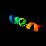
| 1 |
|
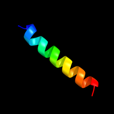
PDB 2zt9 chain F
Region: 29 - 51
Aligned: 23
Modelled: 23
Confidence: 34.8%
Identity: 30%
PDB header:photosynthesis
Chain: F: PDB Molecule:cytochrome b6-f complex subunit 7;
PDBTitle: crystal structure of the cytochrome b6f complex from nostoc sp. pcc2 7120
Phyre2
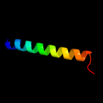
| 2 |
|
PDB 2e74 chain F domain 1
Region: 29 - 51
Aligned: 23
Modelled: 23
Confidence: 34.7%
Identity: 30%
Fold: Single transmembrane helix
Superfamily: PetM subunit of the cytochrome b6f complex
Family: PetM subunit of the cytochrome b6f complex
Phyre2
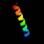
| 3 |
|
PDB 2cpb chain A
Region: 25 - 49
Aligned: 25
Modelled: 25
Confidence: 21.4%
Identity: 28%
PDB header:viral protein
Chain: A: PDB Molecule:m13 major coat protein;
PDBTitle: solution nmr structures of the major coat protein of2 filamentous bacteriophage m13 solubilized in3 dodecylphosphocholine micelles, 25 lowest energy structures
Phyre2
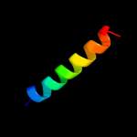
| 4 |
|
PDB 2axt chain E domain 1
Region: 77 - 96
Aligned: 20
Modelled: 20
Confidence: 20.4%
Identity: 40%
Fold: Single transmembrane helix
Superfamily: Cytochrome b559 subunits
Family: Cytochrome b559 subunits
Phyre2
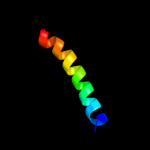
| 5 |
|
PDB 1afo chain B
Region: 82 - 111
Aligned: 30
Modelled: 30
Confidence: 9.7%
Identity: 23%
PDB header:integral membrane protein
Chain: B: PDB Molecule:glycophorin a;
PDBTitle: dimeric transmembrane domain of human glycophorin a, nmr,2 20 structures
Phyre2
|
| Detailed template information | |
Due to computational demand, binding site predictions are not run for batch jobs
If you want to predict binding sites, please manually submit your model of choice to 3DLigandSite
Phyre is for academic use only
| Please cite: Protein structure prediction on
the web: a case study using the Phyre server |
| Kelley LA and Sternberg MJE. Nature Protocols
4, 363 - 371 (2009) [pdf] [Import into BibTeX] |
| |
| If you use the binding site
predictions from 3DLigandSite, please also cite: |
| 3DLigandSite: predicting ligand-binding sites using similar structures. |
| Wass MN, Kelley LA and Sternberg
MJ Nucleic Acids Research 38, W469-73 (2010) [PubMed] |
| |
|
|
|
|