| Secondary structure and disorder prediction | |
| | |
1 | . | . | . | . | . | . | . | . | 10 | . | . | . | . | . | . | . | . | . | 20 | . | . | . | . | . | . | . | . | . | 30 | . | . | . | . | . | . | . | . | . | 40 | . | . |
| Sequence | |
M | S | K | R | M | H | S | H | S | I | A | W | R | K | R | V | I | D | K | A | I | I | V | L | G | A | L | I | A | L | L | E | L | I | R | F | L | L | Q | L | L | N |
| Secondary structure | |
|  | | | | | | | | | | | | | | | | | | | | | | | | | | | | | | | | | | | | | | |
|
|
| SS confidence | |
|
|
|
|
|
|
|
|
|
|
|
|
|
|
|
|
|
|
|
|
|
|
|
|
|
|
|
|
|
|
|
|
|
|
|
|
|
|
|
|
|
|
| Disorder | |
? | ? | ? | ? |
|
|
|
|
|
|
|
|
|
|
|
|
|
|
|
|
|
|
|
|
|
|
|
|
|
|
|
|
|
|
|
|
|
|
|
| ? | ? |
| Disorder confidence | |
|
|
|
|
|
|
|
|
|
|
|
|
|
|
|
|
|
|
|
|
|
|
|
|
|
|
|
|
|
|
|
|
|
|
|
|
|
|
|
|
|
|
| |
| Confidence Key |
| High(9) | |
|
|
|
|
|
|
|
|
|
Low (0) |
| ? | Disordered |
| Alpha helix |
  | Beta strand |
Hover over an aligned region to see model and summary info
Please note, only up to the top 20 hits are modelled to reduce computer load
|
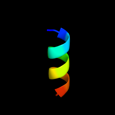
| 1 |
|
PDB 2b2a chain A
Region: 14 - 25
Aligned: 12
Modelled: 12
Confidence: 18.5%
Identity: 67%
PDB header:transferase
Chain: A: PDB Molecule:telomerase reverse transcriptase;
PDBTitle: crystal structure of the ten domain of the telomerase2 reverse transcriptase
Phyre2
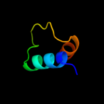
| 2 |
|
PDB 2afv chain B
Region: 1 - 35
Aligned: 35
Modelled: 35
Confidence: 9.9%
Identity: 31%
PDB header:isomerase
Chain: B: PDB Molecule:cobalamin biosynthesis precorrin isomerase;
PDBTitle: the crystal structure of putative precorrin isomerase cbic2 in cobalamin biosynthesis
Phyre2
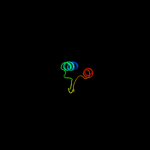
| 3 |
|
PDB 1v9c chain A
Region: 1 - 35
Aligned: 35
Modelled: 35
Confidence: 9.8%
Identity: 37%
Fold: Flavodoxin-like
Superfamily: Precorrin-8X methylmutase CbiC/CobH
Family: Precorrin-8X methylmutase CbiC/CobH
Phyre2
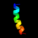
| 4 |
|
PDB 2klu chain A
Region: 20 - 38
Aligned: 19
Modelled: 19
Confidence: 8.5%
Identity: 42%
PDB header:immune system, membrane protein
Chain: A: PDB Molecule:t-cell surface glycoprotein cd4;
PDBTitle: nmr structure of the transmembrane and cytoplasmic domains2 of human cd4
Phyre2
| 5 |
|
PDB 2auh chain B
Region: 11 - 15
Aligned: 5
Modelled: 5
Confidence: 6.5%
Identity: 80%
PDB header:transferase/signaling protein
Chain: B: PDB Molecule:growth factor receptor-bound protein 14;
PDBTitle: crystal structure of the grb14 bps region in complex with2 the insulin receptor tyrosine kinase
Phyre2
|
| Detailed template information | |
Due to computational demand, binding site predictions are not run for batch jobs
If you want to predict binding sites, please manually submit your model of choice to 3DLigandSite
Phyre is for academic use only
| Please cite: Protein structure prediction on
the web: a case study using the Phyre server |
| Kelley LA and Sternberg MJE. Nature Protocols
4, 363 - 371 (2009) [pdf] [Import into BibTeX] |
| |
| If you use the binding site
predictions from 3DLigandSite, please also cite: |
| 3DLigandSite: predicting ligand-binding sites using similar structures. |
| Wass MN, Kelley LA and Sternberg
MJ Nucleic Acids Research 38, W469-73 (2010) [PubMed] |
| |
|
|
|
|