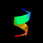
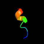
| 1 | c3no7A_ |
|
 |
19.0 |
86 |
PDB header:dna binding protein
Chain: A: PDB Molecule:putative plasmid related protein;
PDBTitle: crystal structure of the centromere-binding protein parb from plasmid2 pcxc100
|
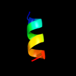
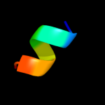
| 2 | c3b8eB_ |
|
 |
10.9 |
42 |
PDB header:hydrolase/transport protein
Chain: B: PDB Molecule:sodium/potassium-transporting atpase subunit
PDBTitle: crystal structure of the sodium-potassium pump
|
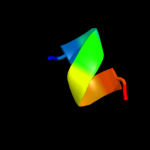
| 3 | d1d4oa_ |
|
 |
10.4 |
50 |
Fold:DHS-like NAD/FAD-binding domain
Superfamily:DHS-like NAD/FAD-binding domain
Family:Transhydrogenase domain III (dIII) |
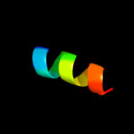
| 4 | d1pnoa_ |
|
 |
9.8 |
60 |
Fold:DHS-like NAD/FAD-binding domain
Superfamily:DHS-like NAD/FAD-binding domain
Family:Transhydrogenase domain III (dIII) |
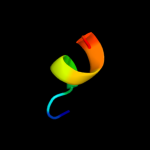
| 5 | c2bruC_ |
|
 |
9.8 |
50 |
PDB header:oxidoreductase
Chain: C: PDB Molecule:nad(p) transhydrogenase subunit beta;
PDBTitle: complex of the domain i and domain iii of escherichia coli2 transhydrogenase
|
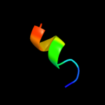
| 6 | c1pt9B_ |
|
 |
9.6 |
50 |
PDB header:oxidoreductase
Chain: B: PDB Molecule:nad(p) transhydrogenase, mitochondrial;
PDBTitle: crystal structure analysis of the diii component of transhydrogenase2 with a thio-nicotinamide nucleotide analogue
|
| 7 | c1erfA_ |
|
 |
6.5 |
75 |
PDB header:viral protein
Chain: A: PDB Molecule:transmembrane glycoprotein;
PDBTitle: conformational mapping of the n-terminal fusion peptide of2 hiv-1 gp41 using 13c-enhanced fourier transform infrared3 spectroscopy (ftir)
|
| 8 | c2pjvA_ |
|
 |
6.4 |
75 |
PDB header:viral protein
Chain: A: PDB Molecule:envelope glycoprotein;
PDBTitle: solution structure of hiv-1 gp41 fusion domain bound to dpc2 micelle
|
| 9 | c3ixzB_ |
|
 |
5.9 |
17 |
PDB header:hydrolase
Chain: B: PDB Molecule:potassium-transporting atpase subunit beta;
PDBTitle: pig gastric h+/k+-atpase complexed with aluminium fluoride
|