| 1 | c3r8t2_ |
|
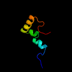 |
99.9 |
100 |
PDB header:ribosome
Chain: 2: PDB Molecule:50s ribosomal protein l34;
PDBTitle: structures of the bacterial ribosome in classical and hybrid states of2 trna binding
|
| 2 | c3i1r2_ |
|
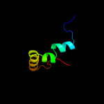 |
99.9 |
100 |
PDB header:ribosome
Chain: 2: PDB Molecule:50s ribosomal protein l34;
PDBTitle: crystal structure of the e. coli 70s ribosome in an2 intermediate state of ratcheting
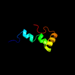
|
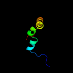
| 3 | c3r8s2_ |
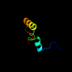
|
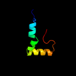 |
99.9 |
100 |
PDB header:ribosome
Chain: 2: PDB Molecule:50s ribosomal protein l34;
PDBTitle: structures of the bacterial ribosome in classical and hybrid states of2 trna binding
|
| 4 | c3izue_ |
|
 |
99.9 |
100 |
PDB header:ribosome
Chain: E: PDB Molecule:50s ribosomal protein l3;
PDBTitle: structural insights into cognate vs. near-cognate discrimination2 during decoding. this entry contains the large subunit of a ribosome3 programmed with a cognate codon
|
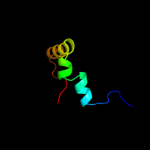
| 5 | c3i202_ |
|
 |
99.9 |
100 |
PDB header:ribosome
Chain: 2: PDB Molecule:50s ribosomal protein l34;
PDBTitle: crystal structure of the e. coli 70s ribosome in an2 intermediate state of ratcheting
|
| 6 | c3izte_ |
|
 |
99.9 |
100 |
PDB header:ribosome
Chain: E: PDB Molecule:50s ribosomal protein l3;
PDBTitle: structural insights into cognate vs. near-cognate discrimination2 during decoding. this entry contains the large subunit of a ribosome3 programmed with a near-cognate codon.
|
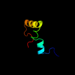
| 7 | c2qbc2_ |
|
 |
99.9 |
100 |
PDB header:ribosome
Chain: 2: PDB Molecule:50s ribosomal protein l34;
PDBTitle: crystal structure of the bacterial ribosome from2 escherichia coli in complex with gentamicin. this file3 contains the 50s subunit of the second 70s ribosome, with4 gentamicin bound. the entire crystal structure contains5 two 70s ribosomes and is described in remark 400.
|
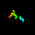
| 8 | c2qao2_ |
|
 |
99.9 |
100 |
PDB header:ribosome
Chain: 2: PDB Molecule:50s ribosomal protein l34;
PDBTitle: crystal structure of the bacterial ribosome from2 escherichia coli in complex with neomycin. this file3 contains the 50s subunit of the second 70s ribosome, with4 neomycin bound. the entire crystal structure contains two5 70s ribosomes and is described in remark 400.
|
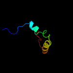
| 9 | c2qba2_ |
|
 |
99.9 |
100 |
PDB header:ribosome
Chain: 2: PDB Molecule:50s ribosomal protein l34;
PDBTitle: crystal structure of the bacterial ribosome from2 escherichia coli in complex with gentamicin. this file3 contains the 50s subunit of the first 70s ribosome, with4 gentamicin bound. the entire crystal structure contains5 two 70s ribosomes and is described in remark 400.
|
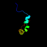
| 10 | c2qam2_ |
|
 |
99.9 |
100 |
PDB header:ribosome
Chain: 2: PDB Molecule:50s ribosomal protein l34;
PDBTitle: crystal structure of the bacterial ribosome from2 escherichia coli in complex with neomycin. this file3 contains the 50s subunit of the first 70s ribosome, with4 neomycin bound. the entire crystal structure contains two5 70s ribosomes and is described in remark 400.
|
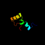
| 11 | c2z4n2_ |
|
 |
99.9 |
100 |
PDB header:ribosome
Chain: 2: PDB Molecule:50s ribosomal protein l34;
PDBTitle: crystal structure of the bacterial ribosome from escherichia2 coli in complex with paromomycin and ribosome recycling3 factor (rrf). this file contains the 50s subunit of the4 second 70s ribosome, with paromomycin and rrf bound. the5 entire crystal structure contains two 70s ribosomes and is6 described in remark 400.
|
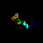
| 12 | c2z4l2_ |
|
 |
99.9 |
100 |
PDB header:ribosome
Chain: 2: PDB Molecule:50s ribosomal protein l34;
PDBTitle: crystal structure of the bacterial ribosome from escherichia2 coli in complex with paromomycin and ribosome recycling3 factor (rrf). this file contains the 50s subunit of the4 first 70s ribosome, with paromomycin and rrf bound. the5 entire crystal structure contains two 70s ribosomes and is6 described in remark 400.
|
| 13 | c3i1n2_ |
|
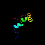 |
99.9 |
100 |
PDB header:ribosome
Chain: 2: PDB Molecule:50s ribosomal protein l34;
PDBTitle: crystal structure of the e. coli 70s ribosome in an2 intermediate state of ratcheting
|
| 14 | c3e1dV_ |
|
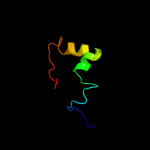 |
99.9 |
100 |
PDB header:ribosome
Chain: V: PDB Molecule:50s ribosomal protein l34;
PDBTitle: structure of the 50s subunit of e. coli ribosome in post-2 accommodation state
|
| 15 | c2qbe2_ |
|
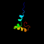 |
99.9 |
100 |
PDB header:ribosome
Chain: 2: PDB Molecule:50s ribosomal protein l34;
PDBTitle: crystal structure of the bacterial ribosome from escherichia2 coli in complex with ribosome recycling factor (rrf). this3 file contains the 50s subunit of the first 70s ribosome,4 with rrf bound. the entire crystal structure contains two5 70s ribosomes and is described in remark 400.
|
| 16 | c2qbg2_ |
|
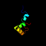 |
99.9 |
100 |
PDB header:ribosome
Chain: 2: PDB Molecule:50s ribosomal protein l34;
PDBTitle: crystal structure of the bacterial ribosome from escherichia2 coli in complex with ribosome recycling factor (rrf). this3 file contains the 50s subunit of the second 70s ribosome,4 with rrf bound. the entire crystal structure contains two5 70s ribosomes and is described in remark 400.
|
| 17 | c2qp12_ |
|
 |
99.9 |
100 |
PDB header:ribosome
Chain: 2: PDB Molecule:50s ribosomal protein l34;
PDBTitle: crystal structure of the bacterial ribosome from escherichia2 coli in complex with spectinomycin and neomycin. this file3 contains the 50s subunit of the second 70s ribosome, with4 neomycin bound. the entire crystal structure contains two5 70s ribosomes.
|
| 18 | c2qoz2_ |
|
 |
99.9 |
100 |
PDB header:ribosome
Chain: 2: PDB Molecule:50s ribosomal protein l34;
PDBTitle: crystal structure of the bacterial ribosome from escherichia2 coli in complex with spectinomycin and neomycin. this file3 contains the 50s subunit of the first 70s ribosome, with4 neomycin bound. the entire crystal structure contains two5 70s ribosomes.
|
| 19 | c2qbi2_ |
|
 |
99.9 |
100 |
PDB header:ribosome
Chain: 2: PDB Molecule:50s ribosomal protein l34;
PDBTitle: crystal structure of the bacterial ribosome from escherichia2 coli in complex with gentamicin and ribosome recycling3 factor (rrf). this file contains the 50s subunit of the4 first 70s ribosome, with gentamicin and rrf bound. the5 entire crystal structure contains two 70s ribosomes and is6 described in remark 400.
|
| 20 | c2qbk2_ |
|
 |
99.9 |
100 |
PDB header:ribosome
Chain: 2: PDB Molecule:50s ribosomal protein l34;
PDBTitle: crystal structure of the bacterial ribosome from escherichia2 coli in complex with gentamicin and ribosome recycling3 factor (rrf). this file contains the 50s subunit of the4 second 70s ribosome, with gentamicin and rrf bound. the5 entire crystal structure contains two 70s ribosomes and is6 described in remark 400.
|
| 21 | c1vs62_ |
|
not modelled |
99.9 |
100 |
PDB header:ribosome
Chain: 2: PDB Molecule:50s ribosomal protein l34;
PDBTitle: crystal structure of the bacterial ribosome from2 escherichia coli in complex with the antibiotic kasugamyin3 at 3.5a resolution. this file contains the 50s subunit of4 one 70s ribosome. the entire crystal structure contains5 two 70s ribosomes and is described in remark 400.
|
| 22 | c2qox2_ |
|
not modelled |
99.9 |
100 |
PDB header:ribosome
Chain: 2: PDB Molecule:50s ribosomal protein l34;
PDBTitle: crystal structure of the bacterial ribosome from escherichia2 coli in complex with spectinomycin. this file contains the3 50s subunit of the second 70s ribosome. the entire crystal4 structure contains two 70s ribosomes.
|
| 23 | c2qov2_ |
|
not modelled |
99.9 |
100 |
PDB header:ribosome
Chain: 2: PDB Molecule:50s ribosomal protein l34;
PDBTitle: crystal structure of the bacterial ribosome from escherichia2 coli in complex with spectinomycin. this file contains the3 50s subunit of the first 70s ribosome. the entire crystal4 structure contains two 70s ribosomes.
|
| 24 | c3df42_ |
|
not modelled |
99.9 |
100 |
PDB header:ribosome
Chain: 2: PDB Molecule:50s ribosomal protein l34;
PDBTitle: crystal structure of the bacterial ribosome from escherichia2 coli in complex with hygromycin b. this file contains the3 50s subunit of the second 70s ribosome. the entire crystal4 structure contains two 70s ribosomes.
|
| 25 | c3df22_ |
|
not modelled |
99.9 |
100 |
PDB header:ribosome
Chain: 2: PDB Molecule:50s ribosomal protein l34;
PDBTitle: crystal structure of the bacterial ribosome from escherichia2 coli in complex with hygromycin b. this file contains the3 50s subunit of the first 70s ribosome. the entire crystal4 structure contains two 70s ribosomes.
|
| 26 | c2aw42_ |
|
not modelled |
99.9 |
100 |
PDB header:ribosome
Chain: 2: PDB Molecule:50s ribosomal protein l34;
PDBTitle: crystal structure of the bacterial ribosome from2 escherichia coli at 3.5 a resolution. this file contains3 the 50s subunit of one 70s ribosome. the entire crystal4 structure contains two 70s ribosomes and is described in5 remark 400.
|
| 27 | c3bbx2_ |
|
not modelled |
99.9 |
100 |
PDB header:ribosome
Chain: 2: PDB Molecule:50s ribosomal protein l34;
PDBTitle: the hsp15 protein fitted into the low resolution cryo-em map of the2 50s.nc-trna.hsp15 complex
|
| 28 | c3e1bV_ |
|
not modelled |
99.9 |
100 |
PDB header:ribosome
Chain: V: PDB Molecule:50s ribosomal protein l34;
PDBTitle: structure of the 50s subunit of e. coli ribosome in pre-2 accommodation state
|
| 29 | c2vhm2_ |
|
not modelled |
99.9 |
100 |
PDB header:ribosome
Chain: 2: PDB Molecule:50s ribosomal protein l34;
PDB Fragment:residues 2-142;
PDBTitle: structure of pdf binding helix in complex with the ribosome2 (part 1 of 4)
|
| 30 | c2vhn2_ |
|
not modelled |
99.9 |
100 |
PDB header:ribosome
Chain: 2: PDB Molecule:50s ribosomal protein l34;
PDB Fragment:residues 2-142
PDBTitle: structure of pdf binding helix in complex with the ribosome.2 (part 2 of 4)
|
| 31 | c3i1p2_ |
|
not modelled |
99.9 |
100 |
PDB header:ribosome
Chain: 2: PDB Molecule:50s ribosomal protein l34;
PDBTitle: crystal structure of the e. coli 70s ribosome in an2 intermediate state of ratcheting
|
| 32 | c3i1t2_ |
|
not modelled |
99.9 |
100 |
PDB header:ribosome
Chain: 2: PDB Molecule:50s ribosomal protein l34;
PDBTitle: crystal structure of the e. coli 70s ribosome in an2 intermediate state of ratcheting
|
| 33 | c2awb2_ |
|
not modelled |
99.9 |
100 |
PDB header:ribosome
Chain: 2: PDB Molecule:50s ribosomal protein l34;
PDBTitle: crystal structure of the bacterial ribosome from2 escherichia coli at 3.5 a resolution. this file contains3 the 50s subunit of the second 70s ribosome. the entire4 crystal structure contains two 70s ribosomes and is5 described in remark 400.
|
| 34 | c2j282_ |
|
not modelled |
99.9 |
100 |
PDB header:ribosome
Chain: 2: PDB Molecule:50s ribosomal protein l34;
PDBTitle: model of e. coli srp bound to 70s rncs
|
| 35 | c2i2v2_ |
|
not modelled |
99.9 |
100 |
PDB header:ribosome
Chain: 2: PDB Molecule:50s ribosomal protein l34;
PDBTitle: crystal structure of ribosome with messenger rna and the2 anticodon stem-loop of p-site trna. this file contains the3 50s subunit of one 70s ribosome. the entire crystal4 structure contains two 70s ribosomes and is described in5 remark 400.
|
| 36 | c2i2t2_ |
|
not modelled |
99.9 |
100 |
PDB header:ribosome
Chain: 2: PDB Molecule:50s ribosomal protein l34;
PDBTitle: crystal structure of ribosome with messenger rna and the2 anticodon stem-loop of p-site trna. this file contains the3 50s subunit of one 70s ribosome. the entire crystal4 structure contains two 70s ribosomes and is described in5 remark 400.
|
| 37 | c2wwq6_ |
|
not modelled |
99.9 |
100 |
PDB header:ribosome
Chain: 6: PDB Molecule:50s ribosomal protein l34;
PDBTitle: e.coli 70s ribosome stalled during translation of tnac2 leader peptide. this file contains the 50s, the p-site3 trna and the tnac leader peptide (part 2 of 2).
|
| 38 | c3j012_ |
|
not modelled |
99.9 |
100 |
PDB header:ribosome/ribosomal protein
Chain: 2: PDB Molecule:50s ribosomal protein l34;
PDBTitle: structure of the ribosome-secye complex in the membrane environment
|
| 39 | c3i222_ |
|
not modelled |
99.9 |
100 |
PDB header:ribosome
Chain: 2: PDB Molecule:50s ribosomal protein l34;
PDBTitle: crystal structure of the e. coli 70s ribosome in an2 intermediate state of ratcheting
|
| 40 | c2rdo2_ |
|
not modelled |
99.9 |
100 |
PDB header:ribosome
Chain: 2: PDB Molecule:50s ribosomal protein l34;
PDBTitle: 50s subunit with ef-g(gdpnp) and rrf bound
|
| 41 | c1vs82_ |
|
not modelled |
99.9 |
100 |
PDB header:ribosome
Chain: 2: PDB Molecule:50s ribosomal protein l34;
PDBTitle: crystal structure of the bacterial ribosome from escherichia coli in2 complex with the antibiotic kasugamyin at 3.5a resolution. this file3 contains the 50s subunit of one 70s ribosome. the entire crystal4 structure contains two 70s ribosomes and is described in remark 400.
|
| 42 | c3fin7_ |
|
not modelled |
99.9 |
64 |
PDB header:ribosome
Chain: 7: PDB Molecule:50s ribosomal protein l34;
PDBTitle: t. thermophilus 70s ribosome in complex with mrna, trnas2 and ef-tu.gdp.kirromycin ternary complex, fitted to a 6.43 a cryo-em map. this file contains the 50s subunit.
|
| 43 | c1sm12_ |
|
not modelled |
99.9 |
70 |
PDB header:ribosome/antibiotic
Chain: 2: PDB Molecule:50s ribosomal protein l34;
PDBTitle: complex of the large ribosomal subunit from deinococcus radiodurans2 with quinupristin and dalfopristin
|
| 44 | c3orb2_ |
|
not modelled |
99.9 |
100 |
PDB header:ribosome
Chain: 2: PDB Molecule:50s ribosomal protein l34;
PDBTitle: crystal structure of the e. coli ribosome bound to cem-101. this file2 contains the 50s subunit of the first 70s ribosome bound to cem-101.
|
| 45 | c3ofc2_ |
|
not modelled |
99.9 |
100 |
PDB header:ribosome
Chain: 2: PDB Molecule:50s ribosomal protein l34;
PDBTitle: crystal structure of the e. coli ribosome bound to chloramphenicol.2 this file contains the 50s subunit of the first 70s ribosome with3 chloramphenicol bound.
|
| 46 | c3ofz2_ |
|
not modelled |
99.9 |
100 |
PDB header:ribosome
Chain: 2: PDB Molecule:50s ribosomal protein l34;
PDBTitle: crystal structure of the e. coli ribosome bound to clindamycin. this2 file contains the 50s subunit of the first 70s ribosome bound to3 clindamycin.
|
| 47 | c3og02_ |
|
not modelled |
99.9 |
100 |
PDB header:ribosome
Chain: 2: PDB Molecule:50s ribosomal protein l34;
PDBTitle: crystal structure of the e. coli ribosome bound to clindamycin. this2 file contains the 50s subunit of the second 70s ribosome.
|
| 48 | c3ofd2_ |
|
not modelled |
99.9 |
100 |
PDB header:ribosome
Chain: 2: PDB Molecule:50s ribosomal protein l34;
PDBTitle: crystal structure of the e. coli ribosome bound to chloramphenicol.2 this file contains the 50s subunit of the second 70s ribosome.
|
| 49 | c1vt22_ |
|
not modelled |
99.9 |
100 |
PDB header:ribosome
Chain: 2: PDB Molecule:50s ribosomal protein l34;
PDBTitle: crystal structure of the e. coli ribosome bound to cem-101. this file2 contains the 50s subunit of the second 70s ribosome.
|
| 50 | c3ofr2_ |
|
not modelled |
99.8 |
100 |
PDB header:ribosome
Chain: 2: PDB Molecule:50s ribosomal protein l34;
PDBTitle: crystal structure of the e. coli ribosome bound to erythromycin. this2 file contains the 50s subunit of the first 70s ribosome with3 erthromycin bound.
|
| 51 | c3oat2_ |
|
not modelled |
99.8 |
100 |
PDB header:ribosome/antibiotic
Chain: 2: PDB Molecule:50s ribosomal protein l34;
PDBTitle: crystal structure of the e. coli ribosome bound to telithromycin. this2 file contains the 50s subunit of the first 70s ribosome with3 telithromycin bound.
|
| 52 | c3ofq2_ |
|
not modelled |
99.8 |
100 |
PDB header:ribosome
Chain: 2: PDB Molecule:50s ribosomal protein l34;
PDBTitle: crystal structure of the e. coli ribosome bound to erythromycin. this2 file contains the 50s subunit of the second 70s ribosome.
|
| 53 | c3oas2_ |
|
not modelled |
99.8 |
100 |
PDB header:ribosome/antibiotic
Chain: 2: PDB Molecule:50s ribosomal protein l34;
PDBTitle: crystal structure of the e. coli ribosome bound to telithromycin. this2 file contains the 50s subunit of the second 70s ribosome.
|
| 54 | c2ftcQ_ |
|
not modelled |
99.7 |
47 |
PDB header:ribosome
Chain: Q: PDB Molecule:39s ribosomal protein l34, mitochondrial;
PDBTitle: structural model for the large subunit of the mammalian mitochondrial2 ribosome
|
| 55 | c3bbo4_ |
|
not modelled |
98.8 |
57 |
PDB header:ribosome
Chain: 4: PDB Molecule:ribosomal protein l34;
PDBTitle: homology model for the spinach chloroplast 50s subunit2 fitted to 9.4a cryo-em map of the 70s chlororibosome
|
| 56 | c2x5aT_ |
|
not modelled |
8.1 |
63 |
PDB header:viral protein
Chain: T: PDB Molecule:orf15;
PDBTitle: structure of the phage p2 baseplate in its activated2 conformation with ca (part 2 of 2)
|

