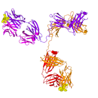
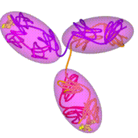
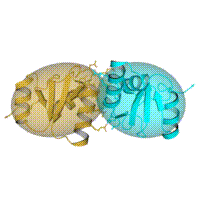
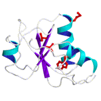
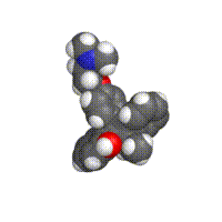
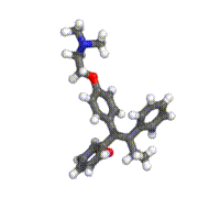
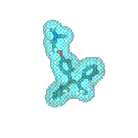
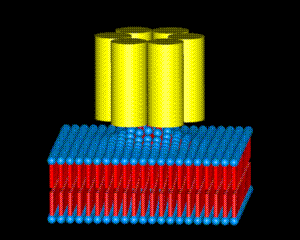
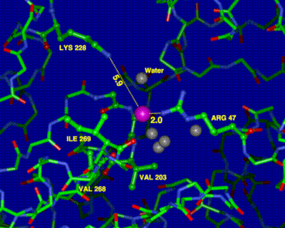
SAMPLE MOLECULAR IMAGES

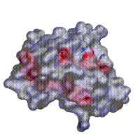
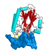
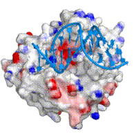
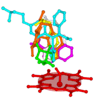
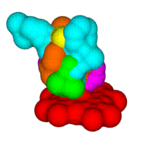
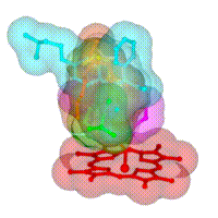
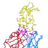
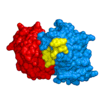
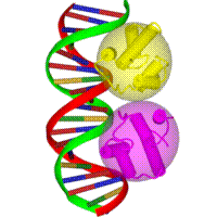
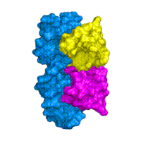
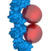
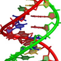
Sample molecular Images produced by the program PREPI (freely available to academic users). The high resolution raytraced versions of these images (TIFF format 2000x2000 pixels) are at the ICRF Anonymous ftp site. To obtain these high resolution images you have three choices:
    (a) Click on the desired image(s) below
    (b) Go directly to the ICRF Anonymous ftp site using your WEB Browser
    (c) Go directly to the ICRF Anonymous ftp site, e.g., using programs FTP, FETCH:
      SITE: ftp.icnet.uk
      LOGIN NAME: ftp
      PASSWORD: your_email_address
      DIRECTORY: /pub/bmm/tmp
If any variations in colour/size are required please contact Suhail A Islam.
An astrix (*) indicates ICRF work.
SM3*:	Breast Tumour-specific Antibody SM3 Complexed with
	its Peptide Epitope (MUC1 antigen). Complex superimposed
	onto 1igt to obtain complete immunugloblin structure
	Dokurno et al, J.MOL.BIOL(1998) 284, 713-728
	(a) SM3 (peptide in yellow) [file 1igt-1.tif]
	(b) Simplified representation (peptide in yellow) [file 1igt-2.tif]
(a) (b)
BRCA1*:	Structure of an XRCC1 BRCT domain and BRCA1 model based upon
	the crystal structure
	Zhang et al, The EMBO Journal Vol. 17, 1998
	(a) XRCC1 BRCT Dimer [file brct-dimer.tif]
	(b) BRCA1 Model [file brca1.tif]
	(c) BRCA1 Model - Charged Surface [file brca1-surf.tif]
(a) (b)
(c)
HAP1*:	The Crystal structure of the human DNA repair endonuclease
	HAP1 suggests the recognition of extra-helical deoxyribose
	at DNA abasic sites. The DNA has been modelled in.
	Gorman et. al., The EMBO Journal Vol.16 No. 21 pp.6548-6558, 1997
	(a)  HAP1 molecule + Modelled DNA [file hap-dna.tif]
	(b)  HAP1 Molecular surface with charge [file hap-dna-1.tif]
(a) (b)
P450*:	MODELLING Cytochrome P450 Substrates
	Scanning for potential substrates for this enzyme - carcinogenic precursors
	(a) Various substrate modeled at Heme catalytic site  [file debris.tif]
	(b) Molecluar Surface of all substrate  [file debris1.tif]
	(c) Transparent Molecular Surface  [file debris2.tif]
	Islam et al,CARCINOGENISIS vol 12,no 12,pp 2211-2219 (1991)
(a) (b)
(c)
1FDL:	COMPLEX (ANTIBODY-ANTIGEN)
	IG*G1 FAB FRAGMENT (ANTI-LYSOZYME ANTIBODY D1.3, KAPPA)
	(a) Molecular Surface [file 1fdl.tif]
	(b) C-alpha with Interacting residues [file 1fdl1.tif]
(a) (b)
1HIM:	IGG2A FAB FRAGMENT (FAB 17/9) COMPLEX WITH A SYNTHETIC
	PEPTIDE FROM INFLUENZA VIRUS HEMAGGLUTININ
	(a) Molecular Surface (peptide in yellow) [file 1him.tif]
(a)
3CRO:	GENE REGULATING PROTEIN / DNA COMPLEX
	434 CRO PROTEIN COMPLEX WITH 20 BASE PAIR DNA
	(a) Ribbon Diagram [file 3cro-1.tif]
	(b) Molecular Surface [file 3cro-surf.tif]
	(c) Protein shown as solid ellipsoids [file 3cro-fancy-1.tif]
(a) (b)
(c)
6BNA:	DEOXYRIBONUCLEIC ACID B-DNA 
	(a) Close up showing base pairing [file 6bna.tif]
(a)
TAMOX:	Crystallographic structure of Tamoxifen
	(a) Sphere representation [file tamox1.tif]
	(b) Bond representation [file tamox1.tif]
	(c) Molecular Surface [file tamox3.tif]
(a) (b)
(c)
Model of Protein-Vesicle "complex". Illustration purpose only
Model of Protein-Lipid "pre-complex". Illustration purpose only
Pt(CN)42- binding in Aldose dehydrogenase (8ADH)